Pylorus

Last updated • 3 min readFrom Wikipedia, The Free Encyclopedia
Pylorus
Gray1050.png
Inside of the stomach (pylorus labeled at center left)
Details
SynonymPyloric region, pyloric part
Identifiers
Latin pylorus
Greek πυλωρός
MeSH D011708
TA98 A05.5.01.017
TA2 2930
FMA 14581
Anatomical terminology

The pylorus ( /pˈlɔːrəs/ or /pɪˈlrəs/ ) connects the stomach to the duodenum. The pylorus is considered as having two parts, the pyloric antrum (opening to the body of the stomach) and the pyloric canal (opening to the duodenum). The pyloric canal ends as the pyloric orifice, which marks the junction between the stomach and the duodenum. The orifice is surrounded by a sphincter, a band of muscle, called the pyloric sphincter. The word pylorus comes from Greek πυλωρός, via Latin. The word pylorus in Greek means "gatekeeper", related to "gate" (Greek : pyle) and is thus linguistically related to the word "pylon". [1]

Contents

Structure

Diagram from cancer.gov:
1. Body of stomach
2. Fundus
3. Anterior wall
4. Greater curvature
5. Lesser curvature
6. Cardia
9. Pyloric sphincter
10. Pyloric antrum
11. Pyloric canal
12. Angular incisure
13. Gastric canal
14. Rugal folds Illu stomach.jpg
Diagram from cancer.gov:
1. Body of stomach
2. Fundus
3. Anterior wall
4. Greater curvature
5. Lesser curvature
6. Cardia
9. Pyloric sphincter
10. Pyloric antrum
11. Pyloric canal
12. Angular incisure
13. Gastric canal
14. Rugal folds

The pylorus is the furthest part of the stomach that connects to the duodenum. It is divided into two parts, the antrum, which connects to the body of the stomach, and the pyloric canal, which connects to the duodenum. [2]

Antrum

The antrum also called the gastric antrum or the pyloric antrum is the initial portion of the pyloric region. It is near the bottom of the stomach, proximal to the pyloric sphincter, which separates the stomach and the duodenum. It may temporarily become partially or completely shut off from the remainder of the stomach during digestion by peristaltic contraction of the prepyloric sphincter; it is demarcated, sometimes, from the pyloric canal by a slight groove.

Canal

The pyloric canal (Latin : canalis pyloricus) is the opening between the stomach and the duodenum. [3] The wall thickness of the pyloric canal is up to 3 millimeters (mm) in infants younger than 30 days, [4] and up to 8 mm in adults. [5]

Sphincter

The pyloric sphincter, surrounding the pyloric orifice is a strong ring of smooth muscle at the end of the pyloric canal which lets food pass from the stomach to the duodenum. It acts as a valve, controlling the outflow of gastric contents into the duodenum [6] and release of chyme. Its function is modulated by both the central and enteric nervous systems. [7] It receives sympathetic innervation from the celiac ganglion.

Histology

Microscopic cross-section of the pylorus Pyloric stomach LPO.JPG
Microscopic cross-section of the pylorus

Under microscopy, the pylorus contains numerous glands, including gastric pits, which constitute about half the depth of the pyloric mucosa. They consist of two or three short closed tubes opening into a common duct or mouth. These tubes are wavy, and are about one-half the length of the duct. The duct is lined by columnar cells, continuous with the epithelium lining the surface of the mucous membrane of the stomach, the tubes by shorter and more cubical cell which are finely granular. The glands contain mucous cells and G cells that secrete gastrin. [8]

The pylorus also contains scattered parietal cells and neuroendocrine cells. These endocrine cells include D cells, which release somatostatin, [9] responsible for shutting off acid secretion. (There is a second hormone-sensitive population near the fundus.) Unstriated muscles, which are entirely involuntary, are located at the pylorus.

Function

The pylorus is one component of the gastrointestinal tract. Food from the stomach, as chyme, passes through the pylorus to the duodenum. The pylorus, through the pyloric sphincter, regulates entry of food from the stomach into the duodenum.

Clinical significance

In such conditions as stomach cancer, tumours may partly block the pyloric canal. A special tube can be implanted surgically to connect the stomach to the duodenum so as to facilitate the passage of food from one to the other. The surgery to place this tube is called a gastroduodenostomy.

Stenosis

Pyloric stenosis refers to a pylorus that is narrow. This is due to congenital hypertrophy of the pyloric sphincter. The lumen of the pylorus is narrower, and less food is able to pass through. This problem is often detected in the early weeks of life. When it is present, a newborn baby may projectile vomit after eating, but despite vomiting remain hungry. Pyloric stenosis may be managed by the insertion of a stent, or through surgical cutting of the pyloric sphincter, a pyloromyotomy. [10]

Other

Additional images

See also

Related Research Articles

<span class="mw-page-title-main">Stomach</span> Digestive organ

The stomach is a muscular, hollow organ in the upper gastrointestinal tract of humans and many other animals, including several invertebrates. The stomach has a dilated structure and functions as a vital organ in the digestive system. The stomach is involved in the gastric phase of digestion, following the cephalic phase in which the sight and smell of food and the act of chewing are stimuli. In the stomach a chemical breakdown of food takes place by means of secreted digestive enzymes and gastric acid.

<span class="mw-page-title-main">Ileum</span> Final section of the small intestine

The ileum is the final section of the small intestine in most higher vertebrates, including mammals, reptiles, and birds. In fish, the divisions of the small intestine are not as clear and the terms posterior intestine or distal intestine may be used instead of ileum. Its main function is to absorb vitamin B12, bile salts, and whatever products of digestion that were not absorbed by the jejunum.

<span class="mw-page-title-main">Small intestine</span> Organ in the gastrointestinal tract

The small intestine or small bowel is an organ in the gastrointestinal tract where most of the absorption of nutrients from food takes place. It lies between the stomach and large intestine, and receives bile and pancreatic juice through the pancreatic duct to aid in digestion. The small intestine is about 5.5 metres long and folds many times to fit in the abdomen. Although it is longer than the large intestine, it is called the small intestine because it is narrower in diameter.

<span class="mw-page-title-main">Peristalsis</span> Radially symmetrical contraction and relaxation of muscles

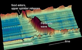
Peristalsis is a type of intestinal motility, characterized by radially symmetrical contraction and relaxation of muscles that propagate in a wave down a tube, in an anterograde direction. Peristalsis is progression of coordinated contraction of involuntary circular muscles, which is preceded by a simultaneous contraction of the longitudinal muscle and relaxation of the circular muscle in the lining of the gut.

Chyme or chymus is the semi-fluid mass of partly digested food that is expelled by the stomach, through the pyloric valve, into the duodenum.

<span class="mw-page-title-main">Esophagogastroduodenoscopy</span> Diagnostic endoscopic procedure

Esophagogastroduodenoscopy (EGD) or oesophagogastroduodenoscopy (OGD), also called by various other names, is a diagnostic endoscopic procedure that visualizes the upper part of the gastrointestinal tract down to the duodenum. It is considered a minimally invasive procedure since it does not require an incision into one of the major body cavities and does not require any significant recovery after the procedure. However, a sore throat is common.

<span class="mw-page-title-main">Pyloric stenosis</span> Medical condition

Pyloric stenosis is a narrowing of the opening from the stomach to the first part of the small intestine. Symptoms include projectile vomiting without the presence of bile. This most often occurs after the baby is fed. The typical age that symptoms become obvious is two to twelve weeks old.

<span class="mw-page-title-main">Brunner's glands</span> Duodenal submucosal cells secreting bicarbonate-rich mucus

Brunner's glands are compound tubuloalveolar submucosal glands found in that portion of the duodenum proximal to the hepatopancreatic sphincter.

<span class="mw-page-title-main">Digestive enzyme</span> Class of enzymes

Digestive enzymes take part in the chemical process of digestion, which follows the mechanical process of digestion. Food consists of macromolecules of proteins, carbohydrates, and fats that need to be broken down chemically by digestive enzymes in the mouth, stomach, pancreas, and duodenum, before being able to be absorbed into the bloodstream. Initial breakdown is achieved by chewing (mastication) and the use of digestive enzymes of saliva. Once in the stomach further mechanical churning takes place mixing the food with secreted gastric acid. Digestive gastric enzymes take part in some of the chemical process needed for absorption. Most of the enzymatic activity, and hence absorption takes place in the duodenum.

<span class="mw-page-title-main">Common hepatic artery</span> Artery

The common hepatic artery is a short blood vessel that supplies oxygenated blood to the liver, pylorus of the stomach, duodenum, pancreas, and gallbladder.

<span class="mw-page-title-main">Foregut</span> Anterior part of the gastrointestinal tract

The foregut in humans is the anterior part of the alimentary canal, from the distal esophagus to the first half of the duodenum, at the entrance of the bile duct. Beyond the stomach, the foregut is attached to the abdominal walls by mesentery. The foregut arises from the endoderm, developing from the folding primitive gut, and is developmentally distinct from the midgut and hindgut. Although the term “foregut” is typically used in reference to the anterior section of the primitive gut, components of the adult gut can also be described with this designation. Pain in the epigastric region, just below the intersection of the ribs, typically refers to structures in the adult foregut.

<span class="mw-page-title-main">Gastric glands</span> Glands in lining of the human stomach

Gastric glands are glands in the lining of the stomach that play an essential role in the process of digestion. Their secretions make up the digestive gastric juice. The gastric glands open into gastric pits in the mucosa. The gastric mucosa is covered in surface mucous cells that produce the mucus necessary to protect the stomach's epithelial lining from gastric acid secreted by parietal cells in the glands, and from pepsin, a secreted digestive enzyme. Surface mucous cells follow the indentations and partly line the gastric pits. Other mucus secreting cells are found in the necks of the glands. These are mucous neck cells that produce a different kind of mucus.

<span class="mw-page-title-main">Gastric outlet obstruction</span> Medical condition

Gastric outlet obstruction (GOO) is a medical condition where there is an obstruction at the level of the pylorus, which is the outlet of the stomach. Individuals with gastric outlet obstruction will often have recurrent vomiting of food that has accumulated in the stomach, but which cannot pass into the small intestine due to the obstruction. The stomach often dilates to accommodate food intake and secretions. Causes of gastric outlet obstruction include both benign causes, such as peptic ulcer disease affecting the area around the pylorus, and malignant causes, such as gastric cancer.

<span class="mw-page-title-main">Gastric mucosa</span> Mucous membrane layer of the stomach

The gastric mucosa is the mucous membrane layer of the stomach, which contains the gastric pits, to which the gastric glands empty. In humans, it is about one mm thick, and its surface is smooth, soft, and velvety. It consists of simple secretory columnar epithelium, an underlying supportive layer of loose connective tissue called the lamina propria, and the muscularis mucosae, a thin layer of muscle that separates the mucosa from the underlying submucosa.

<span class="mw-page-title-main">Curvatures of the stomach</span> Anatomy of the human stomach

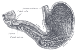
The curvatures of the stomach are the long, convex, lateral surface, and the shorter, concave, medial surface of the stomach, which are referred to as the greater and lesser curvatures, respectively. The greater curvature, which begins at the cardiac notch, and arches backwards, passing inferiorly to the left, is four or five times longer than the lesser curvature, which attaches to the hepatogastric ligament and is supplied by the left gastric artery and right gastric branch of the hepatic artery.

Pyloromyotomy is a surgical procedure in which a portion of the muscle fibers of the pyloric muscle are cut. This is typically done in cases where the contents from the stomach are inappropriately stopped by the pyloric muscle, causing the stomach contents to build up in the stomach and unable to be appropriately digested. The procedure is typically performed in cases of "hypertrophic pyloric stenosis" in young children. In most cases, the procedure can be performed with either an open approach or a laparoscopic approach and the patients typically have good outcomes with minimal complications.

The nervous system, and endocrine system collaborate in the digestive system to control gastric secretions, and motility associated with the movement of food throughout the gastrointestinal tract, including peristalsis, and segmentation contractions.

<span class="mw-page-title-main">Gastrointestinal wall</span> Digestive system structure

The gastrointestinal wall of the gastrointestinal tract is made up of four layers of specialised tissue. From the inner cavity of the gut outwards, these are the mucosa, the submucosa, the muscular layer and the serosa or adventitia.

<span class="mw-page-title-main">Human digestive system</span> Digestive system in humans

The human digestive system consists of the gastrointestinal tract plus the accessory organs of digestion. Digestion involves the breakdown of food into smaller and smaller components, until they can be absorbed and assimilated into the body. The process of digestion has three stages: the cephalic phase, the gastric phase, and the intestinal phase.

References

  1. Harper, Douglas. "Pylorus". Etymology Online. Retrieved 27 March 2014.
  2. Drake, Richard L.; Vogl, Wayne; Tibbitts, Adam W.M. Mitchell; illustrations by Richard; Richardson, Paul (2005). Gray's anatomy for students. Philadelphia: Elsevier/Churchill Livingstone. p. 272. ISBN   978-0-8089-2306-0.
  3. University of Illinois Medical Center:Health Library Archived 2012-04-26 at the Wayback Machine
  4. Rohrschneider, WK; Mittnacht, H; Darge, K; Tröger, J (June 1998). "Pyloric muscle in asymptomatic infants: sonographic evaluation and discrimination from idiopathic hypertrophic pyloric stenosis". Pediatric Radiology. 28 (6): 429–34. doi:10.1007/s002470050377. PMID   9634457.
  5. Lin, Hsien-Ping; Lin, Yu-Chiang; Kuo, Chen-Yun (2015). "Adult idiopathic hypertrophic pyloric stenosis". Journal of the Formosan Medical Association. 114 (7): 659–662. doi: 10.1016/j.jfma.2012.07.001 . ISSN   0929-6646. PMID   26154756.
  6. Snell, Richard S. (2008). Clinical Anatomy by Regions. Lippincott Williams & Wilkins. p. 220. ISBN   978-0781764049.
  7. Bruce M. Carlson (2018). The Human Body: Linking Structure and Function. Academic Press. pp. 331–355. ISBN   9780128043325. The pyloric sphincter controls emptying of the stomach. Its function is coordinated through actions of the central and enteric nervous systems. As acid stomach contents enter the duodenum, the alkaline secretion of Brunner's glands in the duodenum protects the duodenal lining from ulceration.
  8. Cardiac, fundus and pyloric regions of the stomach, Pyloric region. available from: http://histology.leeds.ac.uk/digestive/cardiac_pyloric.php (Last inspected April 16, 2017)
  9. Deakin, Barbara Young; et al. (2006). Wheater's functional histology : a text and colour atlas (5th ed.). Churchill Livingstone/Elsevier. p. 273. ISBN   978-0-443-068-508.
  10. Clayden, Tom Lissauer, Graham (2007). Illustrated textbook of paediatrics (3rd ed.). Edinburgh; New York: Mosby/Elsevier. pp. 207–208. ISBN   9780723433972.{{cite book}}: CS1 maint: multiple names: authors list (link)
  11. Chen, ZM; Scudiere, JR; Abraham, SC; Montgomery, E (2009). "Pyloric gland adenoma: an entity distinct from gastric foveolar type adenoma". Am J Surg Pathol. 33 (2): 186–193. doi:10.1097/PAS.0b013e31817d7ff4. PMID   18830123.