Parietal cell

Last updated
Parietal cell
Parietal cell.png
A parietal cell.
Control-of-stomach-acid-sec.png
Control of stomach acid
Details
Location Stomach
Function Gastric acid, intrinsic factor secretion
Identifiers
Latin exocrinocytus parietalis
MeSH D010295
TH H3.04.02.1.00033
Anatomical terms of microanatomy

Parietal cells (also known as oxyntic cells) are epithelial cells in the stomach that secrete hydrochloric acid (HCl) and intrinsic factor. These cells are located in the gastric glands found in the lining of the fundus and body regions of the stomach. [1] They contain an extensive secretory network of canaliculi from which the HCl is secreted by active transport into the stomach. The enzyme hydrogen potassium ATPase (H+/K+ ATPase) is unique to the parietal cells and transports the H+ against a concentration gradient of about 3 million to 1,[ citation needed ] which is the steepest[ citation needed ] ion gradient formed in the human body. Parietal cells are primarily regulated via histamine, acetylcholine and gastrin signalling from both central and local modulators.

Contents

Structure

Canaliculus

A canaliculus is an adaptation found on gastric parietal cells. It is a deep infolding, or little channel, which serves to increase the surface area, e.g. for secretion. The parietal cell membrane is dynamic; the numbers of canaliculi rise and fall according to secretory need. This is accomplished by the fusion of canalicular precursors, or tubulovesicles, with the membrane to increase surface area, and the reciprocal endocytosis of the canaliculi (reforming the tubulovesicles) to decrease it. [2]

Function

Hydrochloric acid secretion

Hydrochloric acid is formed in the following manner:

Human parietal cells (pink staining) - stomach. Parietal cells.jpg
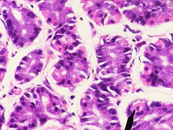
Human parietal cells (pink staining) – stomach.

As a result of the cellular export of hydrogen ions, the gastric lumen is maintained as a highly acidic environment. The acidity aids in digestion of food by promoting the unfolding (or denaturing) of ingested proteins. As proteins unfold, the peptide bonds linking component amino acids are exposed. Gastric HCl simultaneously cleaves pepsinogen, a zymogen, into active pepsin, an endopeptidase that advances the digestive process by breaking the now-exposed peptide bonds, a process known as proteolysis.

Regulation

Parietal cells secrete acid in response to three types of stimuli: [3]

Activation of histamine through H2 receptor causes increases in the intracellular cAMP level while ACh through M3 receptor and gastrin through CCK2 receptor increases intracellular calcium level. These receptors are present on basolateral side of membrane.

Increased cAMP level results in increased protein kinase A. Protein kinase A phosphorylates proteins involved in the transport of H+/K+-ATPase from the cytoplasm to the cell membrane. This causes resorption of K+ ions and secretion of H+ ions. The pH of the secreted fluid can fall by 0.8.

Gastrin primarily induces acid-secretion indirectly, increasing histamine synthesis in ECL cells, which in turn signal parietal cells via histamine release and H2 stimulation. [5] Gastrin itself has no effect on the maximum histamine-stimulated gastric acid secretion. [6]

The effect of histamine, acetylcholine and gastrin is synergistic, that is, effect of two simultaneously is more than additive of effect of the two individually. It helps in non-linear increase of secretion with stimuli physiologically. [7]

Intrinsic factor secretion

Parietal cells also produce a glycoprotein known as intrinsic factor. Intrinsic factor is required for the absorption of vitamin B12 in the diet. A long-term deficiency in vitamin B12 can lead to megaloblastic anemia, characterized by large fragile red blood cells. Pernicious anaemia results from autoimmune destruction of gastric parietal cells, precluding the synthesis of intrinsic factor and, by extension, absorption of vitamin B12. Pernicious anemia also leads to megaloblastic anemia. Atrophic gastritis, particularly in the elderly, will cause an inability to absorb B12 and can lead to deficiencies such as decreased DNA synthesis and nucleotide metabolism in the bone marrow.

Clinical significance

Immunofluorescence staining pattern of gastric parietal antibodies on a stomach section GASTRIC PARIETAL CELL ANTIBODIES.jpg
Immunofluorescence staining pattern of gastric parietal antibodies on a stomach section
Parietal cells are part of fundic gland polyps (here shown in high magnification). Histopathology of fundic gland polyp, high magnification, annotated.jpg
Parietal cells are part of fundic gland polyps (here shown in high magnification).

See also

Related Research Articles

Digestion is the breakdown of large insoluble food compounds into small water-soluble components so that they can be absorbed into the blood plasma. In certain organisms, these smaller substances are absorbed through the small intestine into the blood stream. Digestion is a form of catabolism that is often divided into two processes based on how food is broken down: mechanical and chemical digestion. The term mechanical digestion refers to the physical breakdown of large pieces of food into smaller pieces which can subsequently be accessed by digestive enzymes. Mechanical digestion takes place in the mouth through mastication and in the small intestine through segmentation contractions. In chemical digestion, enzymes break down food into the small compounds that the body can use.

<span class="mw-page-title-main">Delta cell</span>

Delta cells are somatostatin-producing cells. They can be found in the stomach, intestine and the pancreatic islets. Delta cells comprise ca 5% of the cells in the islets but may interact with many more islet cells than suggested by their low numbers. In rodents, delta-cells are located in the periphery of the islets; in humans the islet architecture is generally less organized and delta-cells are frequently observed inside the islets as well. In both species, the peptide hormone Urocortin III (Ucn3) is a major local signal that is released from beta cells to induce the local secretion of somatostatin. It has also been suggested that somatostatin may be implicated in insulin-induced hypoglycaemia through a mechanism involving SGLT-2 receptors. Ghrelin can also strongly stimulate somatostatin secretion, thus indirectly inhibiting insulin release. Viewed under an electron microscope, delta-cells can be identified as cells with smaller and slightly more compact granules than beta cells.

<span class="mw-page-title-main">Intrinsic factor</span> Glycoprotein produced in the stomach which binds to vitamin B12

Intrinsic factor (IF), cobalamin binding intrinsic factor, also known as gastric intrinsic factor (GIF), is a glycoprotein produced by the parietal cells (in humans) or chief cells (in rodents) of the stomach. It is necessary for the absorption of vitamin B12 later on in the distal ileum of the small intestine. In humans, the gastric intrinsic factor protein is encoded by the CBLIF gene. Haptocorrin (transcobalamin I) is another glycoprotein secreted by the salivary glands which binds to vitamin B12. Vitamin B12 is acid-sensitive and in binding to haptocorrin it can safely pass through the acidic stomach to the duodenum.

H<sub>2</sub> receptor antagonist Class of medications

H2 antagonists, sometimes referred to as H2RAs and also called H2 blockers, are a class of medications that block the action of histamine at the histamine H2 receptors of the parietal cells in the stomach. This decreases the production of stomach acid. H2 antagonists can be used in the treatment of dyspepsia, peptic ulcers and gastroesophageal reflux disease. They have been surpassed by proton pump inhibitors (PPIs). The PPI omeprazole was found to be more effective at both healing and alleviating symptoms of ulcers and reflux oesophagitis than the H2 blockers ranitidine and cimetidine.

<span class="mw-page-title-main">Zollinger–Ellison syndrome</span> Condition in which tumours stimulate excessive gastric acid production

Zollinger–Ellison syndrome is rare disease in which tumors cause the stomach to produce too much acid, resulting in peptic ulcers. Symptoms include abdominal pain and diarrhea.

<span class="mw-page-title-main">Pernicious anemia</span> Anemia caused by vitamin B12 deficiency

Pernicious anemia is a disease where not enough red blood cells are produced due to a deficiency of vitamin B12. Those affected often have a gradual onset. The most common initial symptoms are feeling tired and weak. Other symptoms may include shortness of breath, feeling faint, a smooth red tongue, pale skin, chest pain, nausea and vomiting, loss of appetite, heartburn, numbness in the hands and feet, difficulty walking, memory loss, muscle weakness, poor reflexes, blurred vision, clumsiness, depression, and confusion. Without treatment, some of these problems may become permanent.

<span class="mw-page-title-main">Gastrin</span> Mammalian protein found in Homo sapiens

Gastrin is a peptide hormone that stimulates secretion of gastric acid (HCl) by the parietal cells of the stomach and aids in gastric motility. It is released by G cells in the pyloric antrum of the stomach, duodenum, and the pancreas.

<span class="mw-page-title-main">Gastric acid</span> Digestive fluid formed in the stomach

Gastric acid, gastric juice, or stomach acid is a digestive fluid formed within the stomach lining. With a pH between 1.5 and 3, gastric acid plays a key role in digestion of proteins by activating digestive enzymes, which together break down the long chains of amino acids of proteins. Gastric acid is regulated in feedback systems to increase production when needed, such as after a meal. Other cells in the stomach produce bicarbonate, a base, to buffer the fluid, ensuring a regulated pH. These cells also produce mucus – a viscous barrier to prevent gastric acid from damaging the stomach. The pancreas further produces large amounts of bicarbonate and secretes bicarbonate through the pancreatic duct to the duodenum to neutralize gastric acid passing into the digestive tract.

The histamine receptors are a class of G protein–coupled receptors which bind histamine as their primary endogenous ligand.

<span class="mw-page-title-main">Achlorhydria</span> Medical condition

Achlorhydria and hypochlorhydria refer to states where the production of hydrochloric acid in gastric secretions of the stomach and other digestive organs is absent or low, respectively. It is associated with various other medical problems.

<span class="mw-page-title-main">Digestive enzyme</span> Class of enzymes

Digestive enzymes are a group of enzymes that break down polymeric macromolecules into their smaller building blocks, in order to facilitate their absorption into the cells of the body. Digestive enzymes are found in the digestive tracts of animals and in the tracts of carnivorous plants, where they aid in the digestion of food, as well as inside cells, especially in their lysosomes, where they function to maintain cellular survival. Digestive enzymes of diverse specificities are found in the saliva secreted by the salivary glands, in the secretions of cells lining the stomach, in the pancreatic juice secreted by pancreatic exocrine cells, and in the secretions of cells lining the small and large intestines.

<span class="mw-page-title-main">Atrophic gastritis</span> Medical condition

Atrophic gastritis is a process of chronic inflammation of the gastric mucosa of the stomach, leading to a loss of gastric glandular cells and their eventual replacement by intestinal and fibrous tissues. As a result, the stomach's secretion of essential substances such as hydrochloric acid, pepsin, and intrinsic factor is impaired, leading to digestive problems. The most common are vitamin B12 deficiency possibly leading to pernicious anemia; and malabsorption of iron, leading to iron deficiency anaemia. It can be caused by persistent infection with Helicobacter pylori, or can be autoimmune in origin. Those with autoimmune atrophic gastritis (Type A gastritis) are statistically more likely to develop gastric carcinoma, Hashimoto's thyroiditis, and achlorhydria.

<span class="mw-page-title-main">Enterochromaffin-like cell</span>

Enterochromaffin-like cells or ECL cells are a type of neuroendocrine cell found in the gastric glands of the gastric mucosa beneath the epithelium, in particular in the vicinity of parietal cells, that aid in the production of gastric acid via the release of histamine. They are also considered a type of enteroendocrine cell.

<span class="mw-page-title-main">G cell</span> Type of cell in the stomach and duodenum that secretes gastrin

In anatomy, the G cell or gastrin cell is a type of cell in the stomach and duodenum that secretes gastrin. It works in conjunction with gastric chief cells and parietal cells. G cells are found deep within the pyloric glands of the stomach antrum, and occasionally in the pancreas and duodenum. The vagus nerve innervates the G cells. Gastrin-releasing peptide is released by the post-ganglionic fibers of the vagus nerve onto G cells during parasympathetic stimulation. The peptide hormone bombesin also stimulates gastrin from G cells. Gastrin-releasing peptide, as well as the presence of amino acids in the stomach, stimulates the release of gastrin from the G cells. Gastrin stimulates enterochromaffin-like cells to secrete histamine. Gastrin also targets parietal cells by increasing the amount of histamine and the direct stimulation by gastrin, causing the parietal cells to increase HCl secretion in the stomach. G-cells frequently express PD-L1 during homeostasis which protects them from Helicobacter pylori-induced immune destruction

<span class="mw-page-title-main">Gastrinoma</span> Medical condition

Gastrinomas are neuroendocrine tumors (NETs), usually located in the duodenum or pancreas, that secrete gastrin and cause a clinical syndrome known as Zollinger–Ellison syndrome (ZES). A large number of gastrinomas develop in the pancreas or duodenum, with near-equal frequency, and approximately 10% arise as primary neoplasms in lymph nodes of the pancreaticoduodenal region.

<span class="mw-page-title-main">Gastric glands</span> Glands in lining of the human stomach

The gastric glands are glands in the lining of the stomach that play an essential role in the process of digestion. All of the glands have mucus-secreting foveolar cells. Mucus lines the entire stomach, and protects the stomach lining from the effects of hydrochloric acid released from other cells in the glands.

Gastric hydrogen potassium ATPase, also known as H+/K+ ATPase, is an enzyme which functions to acidify the stomach. It is a member of the P-type ATPases, also known as E1-E2 ATPases due to its two states.

In endocrinology, secretagogue is a substance that causes another substance to be secreted. The word contains the suffix -agogue, which refers to something that leads to something else; a secretagogue thus leads to secretion.

The nervous system, and endocrine system collaborate in the digestive system to control gastric secretions, and motility associated with the movement of food throughout the gastrointestinal tract, including peristalsis, and segmentation contractions.

<span class="mw-page-title-main">Human digestive system</span> Digestive system in humans

The human digestive system consists of the gastrointestinal tract plus the accessory organs of digestion. Digestion involves the breakdown of food into smaller and smaller components, until they can be absorbed and assimilated into the body. The process of digestion has three stages: the cephalic phase, the gastric phase, and the intestinal phase.

References

  1. Hunt, A; Harrington, D; Robinson, S (4 June 2014). "Vitamin B12 deficiency" (PDF). BMJ. 349: g5226. doi:10.1136/bmj.g5226. PMID   25189324. S2CID   28782021. Archived from the original (PDF) on 12 March 2017. Retrieved 9 May 2018. Lock-green.svg
  2. Sahoo, N; Gu, M; Zhang, X (8 May 2017). "Gastric Acid Secretion from Parietal Cells Is Mediated by a Ca(2+) Efflux Channel in the Tubulovesicle". Developmental Cell. 41 (3): 262–273.e6. doi:10.1016/j.devcel.2017.04.003. PMC   5497767 . PMID   28486130.
  3. Boulpaep, Walter (2009). Medical Physiology. Philadelphia: Saunders. pp. 898–899. ISBN   978-1-4160-3115-4.
  4. "Gastric acid secretion - Homo sapiens". KEGG. Retrieved June 1, 2011.
  5. Waldum, Helge L., Kleveland, Per M., et al. (2009)'Interactions between gastric acid secretagogues and the localization of the gastrin receptor', Scandinavian Journal of Gastroenterology, 44:4,390—393.
  6. Kleveland PM, Waldum HL, Larsson M. Gastric acid secretion in the totally isolated, vascularly perfused rat stomach. A selective muscarinic-1 agent does, whereas gastrin does not, augment maximal histamine-stimulated acid secretion. Scandinavian Journal of Gastroenterology, 1987;22:705–713.
  7. Ganong's Review of Medical Physiology 24th edition. Lange.
  8. Naziheh Assarzadegan, M.D., Raul S. Gonzalez, M.D. "Stomach Polyps - Fundic gland polyp". PathologyOutlines.{{cite web}}: CS1 maint: multiple names: authors list (link) Topic Completed: 1 November 2017. Minor changes: 11 December 2019