Abdominopelvic cavity

Last updated
Abdominopelvic cavity
Scheme body cavities-en.svg
Details
Identifiers
Latin cavitas abdominis et pelvis
TA98 A01.1.00.050
A10.1.00.000
TA2 3699
FMA 12267
Anatomical terminology

The abdominopelvic cavity is a body cavity that consists of the abdominal cavity and the pelvic cavity. [1] The upper portion is the abdominal cavity, and it contains the stomach, liver, pancreas, spleen, gallbladder, kidneys, small intestine, and most of the large intestine. The lower portion is the pelvic cavity, and it contains the urinary bladder, the rest of the large intestine (the lower portion), and the internal reproductive organs. [2] [3]

Contents

There is no membrane that separates out the abdominal cavity from the pelvic cavity, so the terms abdominal pelvis and peritoneal cavity are sometimes used.

There are many diseases and disorders associated with the organs of the abdominopelvic cavity.

Structure and physiology

The stomach sits on the left side, which is attached to the esophagus tube. Food comes through the esophagus, goes behind all of the other organs in the thoracic cavity, then comes out through the esophagus and opens up into the stomach. The stomach is more of an acidic environment, and begins the major processes of digestion. The food particles must be broken down before entering the small intestine.

The small intestine is about 20 feet and goes behind the large intestine, and then makes a mass of curly tube. The small intestine is divided into three parts: the duodenum, jejunum, and the ileum. The duodenum receives particles from different organs, like the pancreas. The pancreas produces the hormone insulin, which helps regulate blood sugar. [4] [5] The second part is the jejunum, which is located in the middle of the small intestine. The final part of the small intestine is the ilium. It is connected to the large intestine. The ileum is connected to the ileum cecum valve, which is the beginning of the large intestine. The large intestine, also known as the large bowel, is the last part of the gastrointestinal (GI) tract and of the digestive system.

The liver lies in the right hypochondrium area and the greater portion of the epigastric areas of the abdominal cavity under the diaphragm within the rib cage. [6] It is a processing and detoxifying organ. [7] It filters blood and eliminates wastes and toxins from the bloodstream. [8]

The gallbladder is located on the undersurface of the right lobe of the liver. [9] It produces bile, which is used to help process fats in the body. [9] Humans can live without the gallbladder. [10]

The largest lymphatic organ is the spleen, which is dark purple and located under the lower ribs on the left side of the upper abdomen. [11] [12] It filters out the red blood cells and pulls out the old blood cells and cleans them out. [11] [12]

Coming off the cecum (the tiny tail piece) is the appendix. It is a small organ attached to the large intestine in the lower right side of the abdomen. Anatomists and medical professionals have traditionally considered the appendix a vestigial organ. Later research suggests that it may have an immunological function. [13]

On the very dorsal aspect of the abdominal cavity, there are two bean-shaped organs. These are the left and right kidneys. The kidneys filter the blood and urine.

The urinary tract can be divided into the upper urinary tract and the lower urinary tract. The upper urinary tract consists of the kidneys and the ureters, and the lower urinary tract consists of the bladder and the urethra. [14]

Clinical significance

The stomach can be affected by diseases. Helicobacter pylori, previously known as Campylobacter pylori, is a Gram-negative, microaerophilic bacterium infection usually found in the stomach. [15] People who are infected with Campylobacter pylori should drink extra fluids as long as the diarrhea lasts. Antibiotics are needed or prescribed to people who are very ill or at high risk for severe disease, such as people with severely weakened immune systems. This includes people with HIV/AIDS, those receiving chemotherapy, and those with the blood disorders thalassemia and hypogammaglobulinemia.

One disease that affects the lining of the GI tract is Crohn's disease, which is a chronic inflammatory condition. [16] Crohn's most commonly affects the end of the small bowel (the ileum) and the beginning of the colon, but it may affect any part of the GI tract. Crohn's disease affects both the large and small intestine.

Infection of the appendix is appendicitis. When there is a buildup of bacteria, the appendix can get inflamed and swollen, and this leads to appendicitis.

Pancreatitis is the inflammation of the pancreas. Acute pancreatitis is sudden inflammation of the pancreas that may be mild or life-threatening but usually subsides. Gallstones and excessive alcohol use are the main causes of acute pancreatitis. Severe abdominal pain is the predominant symptom.

A person can puncture their spleen if they crack their rib. Because there is a strong flow of blood in the spleen, there is a higher chance of excessive bleeding.

Cirrhosis is a complication of many liver diseases characterized by abnormal structure and function of the liver. The diseases that lead to cirrhosis do so because they injure and kill liver cells, after which the inflammation and repair that is associated with the dying liver cells causes, scar tissue to form. The liver cells that do not die multiply in an attempt to replace the cells that have died. This results in clusters of newly formed liver cells (regenerative nodules) within the scar tissue. A 2008 review states, "The exact prevalence of cirrhosis worldwide is unknown. It was estimated at 0·15% or 400 000 in the USA, 7 which accounted for more than 25000 deaths and 373000 hospital discharges in 1998." [17]

Urinary tract infections are caused by microbes such as bacteria overcoming the body's defenses in the urinary tract. [18] They can affect the kidneys, the bladder, and the tubes that run between them.

A consequence of a fatty diet is diarrhea, due to the fact that the body is no longer processing the food as well.

Additional images

Related Research Articles

<span class="mw-page-title-main">Peritoneum</span> Serous membrane that forms lining of abdominal cavity or coelom

The peritoneum is the serous membrane forming the lining of the abdominal cavity or coelom in amniotes and some invertebrates, such as annelids. It covers most of the intra-abdominal organs, and is composed of a layer of mesothelium supported by a thin layer of connective tissue. This peritoneal lining of the cavity supports many of the abdominal organs and serves as a conduit for their blood vessels, lymphatic vessels, and nerves.

<span class="mw-page-title-main">Abdominal cavity</span> Body cavity in the abdominal area

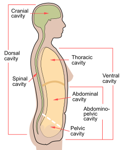
The abdominal cavity is a large body cavity in humans and many other animals that contain organs. It is a part of the abdominopelvic cavity. It is located below the thoracic cavity, and above the pelvic cavity. Its dome-shaped roof is the thoracic diaphragm, a thin sheet of muscle under the lungs, and its floor is the pelvic inlet, opening into the pelvis.

<span class="mw-page-title-main">Gastrointestinal tract</span> Organ system within humans and other animals

The gastrointestinal tract is the tract or passageway of the digestive system that leads from the mouth to the anus. The GI tract contains all the major organs of the digestive system, in humans and other animals, including the esophagus, stomach, and intestines. Food taken in through the mouth is digested to extract nutrients and absorb energy, and the waste expelled at the anus as faeces. Gastrointestinal is an adjective meaning of or pertaining to the stomach and intestines.

<span class="mw-page-title-main">Duodenum</span> First section of the small intestine

The duodenum is the first section of the small intestine in most higher vertebrates, including mammals, reptiles, and birds. In mammals it may be the principal site for iron absorption. The duodenum precedes the jejunum and ileum and is the shortest part of the small intestine.

<span class="mw-page-title-main">Body cavity</span> Internal space within a multicellular organism

A body cavity is any space or compartment, or potential space, in an animal body. Cavities accommodate organs and other structures; cavities as potential spaces contain fluid.

<span class="mw-page-title-main">Small intestine</span> Organ in the gastrointestinal tract

The small intestine or small bowel is an organ in the gastrointestinal tract where most of the absorption of nutrients from food takes place. It lies between the stomach and large intestine, and receives bile and pancreatic juice through the pancreatic duct to aid in digestion. The small intestine is about 5.5 metres long and folds many times to fit in the abdomen. Although it is longer than the large intestine, it is called the small intestine because it is narrower in diameter.

The excretory system is a passive biological system that removes excess, unnecessary materials from the body fluids of an organism, so as to help maintain internal chemical homeostasis and prevent damage to the body. The dual function of excretory systems is the elimination of the waste products of metabolism and to drain the body of used up and broken down components in a liquid and gaseous state. In humans and other amniotes, most of these substances leave the body as urine and to some degree exhalation, mammals also expel them through sweating.

<span class="mw-page-title-main">Organ (biology)</span> Collection of tissues with similar functions

In a multicellular organism, an organ is a collection of tissues joined in a structural unit to serve a common function. In the hierarchy of life, an organ lies between tissue and an organ system. Tissues are formed from same type cells to act together in a function. Tissues of different types combine to form an organ which has a specific function. The intestinal wall for example is formed by epithelial tissue and smooth muscle tissue. Two or more organs working together in the execution of a specific body function form an organ system, also called a biological system or body system.

<span class="mw-page-title-main">Fistula</span> Abnormal connection between two epithelialized surfaces, often organs

In anatomy, a fistula is an abnormal connection joining two hollow spaces, such as blood vessels, intestines, or other hollow organs to each other, often resulting in an abnormal flow of fluid from one space to the other. An anal fistula connects the anal canal to the perianal skin. An anovaginal or rectovaginal fistula is a hole joining the anus or rectum to the vagina. A colovaginal fistula joins the space in the colon to that in the vagina. A urinary tract fistula is an abnormal opening in the urinary tract or an abnormal connection between the urinary tract and another organ. An abnormal communication between the bladder and the uterus is called a vesicouterine fistula, while if it is between the bladder and the vagina it is known as a vesicovaginal fistula, and if between the urethra and the vagina: a urethrovaginal fistula. When occurring between two parts of the intestine, it is known as an enteroenteral fistula, between the small intestine and the skin as an enterocutaneous fistula, and between the colon and the skin as a colocutaneous fistula.

<span class="mw-page-title-main">Portal vein</span> Short thick vein formed by the union of the superior mesenteric vein and the splenic vein

The portal vein or hepatic portal vein (HPV) is a blood vessel that carries blood from the gastrointestinal tract, gallbladder, pancreas and spleen to the liver. This blood contains nutrients and toxins extracted from digested contents. Approximately 75% of total liver blood flow is through the portal vein, with the remainder coming from the hepatic artery proper. The blood leaves the liver to the heart in the hepatic veins.

<span class="mw-page-title-main">Abdominal pain</span> Stomach aches

Abdominal pain, also known as a stomach ache, Is a symptom associated with both non-serious and serious medical issues. Since the abdomen contains most of the body's vital organs, it can be an indicator of a wide variety of diseases. Given that, approaching the examination of a person and planning of a differential diagnosis is extremely important.

<span class="mw-page-title-main">Gastritis</span> Stomach disease that is an inflammation of the lining of the stomach

Gastritis is inflammation of the lining of the stomach. It may occur as a short episode or may be of a long duration. There may be no symptoms but, when symptoms are present, the most common is upper abdominal pain. Other possible symptoms include nausea and vomiting, bloating, loss of appetite and heartburn. Complications may include stomach bleeding, stomach ulcers, and stomach tumors. When due to autoimmune problems, low red blood cells due to not enough vitamin B12 may occur, a condition known as pernicious anemia.

<span class="mw-page-title-main">Gastrointestinal disease</span> Medical condition

Gastrointestinal diseases refer to diseases involving the gastrointestinal tract, namely the esophagus, stomach, small intestine, large intestine and rectum, and the accessory organs of digestion, the liver, gallbladder, and pancreas.

<span class="mw-page-title-main">Abdominal examination</span> Physical examination of abdomen

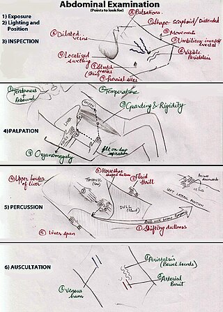
An abdominal examination is a portion of the physical examination which a physician or nurse uses to clinically observe the abdomen of a patient for signs of disease. The abdominal examination is conventionally split into four different stages: first, inspection of the patient and the visible characteristics of their abdomen. Auscultation (listening) of the abdomen with a stethoscope. Palpation of the patient's abdomen. Finally, percussion (tapping) of the patient's abdomen and abdominal organs. Depending on the need to test for specific diseases such as ascites, special tests may be performed as a part of the physical examination. An abdominal examination may be performed because the physician suspects a disease of the organs inside the abdominal cavity (including the liver, spleen, large or small intestines), or simply as a part of a complete physical examination for other conditions. In a complete physical examination, the abdominal exam classically follows the respiratory examination and cardiovascular examination.

<span class="mw-page-title-main">Superior mesenteric vein</span> Vein which drains blood from the small intestine

In human anatomy, the superior mesenteric vein (SMV) is a blood vessel that drains blood from the small intestine. Behind the neck of the pancreas, the superior mesenteric vein combines with the splenic vein to form the portal vein that carries blood to the liver. The superior mesenteric vein lies to the right of the similarly named artery, the superior mesenteric artery, which originates from the abdominal aorta.

<span class="mw-page-title-main">Abdomen</span> Part of the body between the chest and pelvis

The abdomen is the part of the body between the thorax (chest) and pelvis, in humans and in other vertebrates. The abdomen is the front part of the abdominal segment of the torso. The area occupied by the abdomen is called the abdominal cavity. In arthropods, it is the posterior tagma of the body; it follows the thorax or cephalothorax.

<span class="mw-page-title-main">Pelvic cavity</span> Body cavity bounded by the pelvic bones

The pelvic cavity is a body cavity that is bounded by the bones of the pelvis. Its oblique roof is the pelvic inlet. Its lower boundary is the pelvic floor.

<span class="mw-page-title-main">Stomach disease</span> Medical condition

Stomach diseases include gastritis, gastroparesis, Crohn's disease and various cancers.

<span class="mw-page-title-main">Abdominal trauma</span> Medical condition

Abdominal trauma is an injury to the abdomen. Signs and symptoms include abdominal pain, tenderness, rigidity, and bruising of the external abdomen. Complications may include blood loss and infection.

<span class="mw-page-title-main">Human digestive system</span> Digestive system in humans

The human digestive system consists of the gastrointestinal tract plus the accessory organs of digestion. Digestion involves the breakdown of food into smaller and smaller components, until they can be absorbed and assimilated into the body. The process of digestion has three stages: the cephalic phase, the gastric phase, and the intestinal phase.

References

  1. Rollins, Jeannean Hall; Long, Bruce W.; Curtis, Tammy (2022). Merrill's Atlas of Radiographic Positioning and Procedures - 3-Volume Set - E-Book. Elsevier Health Sciences. p. 131. ISBN   978-0-32-383323-3.
  2. Tortora, Gerard J.; Derrickson, Bryan H. (2017). Introduction to the Human Body. John Wiley & Sons. p. 17. ISBN   978-1-11-930666-5.
  3. Cooper, Kim; Gosnell, Kelly (2014). Foundations and Adult Health Nursing. Elsevier Health Sciences. p. 1228. ISBN   978-0-32-310001-4.
  4. Zhang, Liang; Thurber, Greg M. (2017). "Imaging in Diabetes". Imaging and Metabolism. Springer. p. 175. ISBN   978-3-31-961401-4.
  5. Sarafino, Edward P.; Smith, Timothy W.; King, David B.; Longis, Anita De (2020). Health Psychology: Biopsychosocial Interactions. John Wiley & Sons. p. 462. ISBN   978-1-11-950694-2.
  6. Reynolds, James C (2016). The Netter Collection of Medical Illustrations: Digestive System: Part III - Liver, etc. Elsevier Health Sciences. p. 5. ISBN   978-0-32-338934-1.
  7. Patton, Kevin T.; Thibodeau, Gary A. (2018). Anthony's Textbook of Anatomy & Physiology - E-Book. Elsevier Health Sciences. p. 890. ISBN   978-0-32-370930-9.
  8. Noland, Diana; Drisko, Jeanne A.; Uitgever, Leigh Wagner (2020). Integrative and Functional Medical Nutrition Therapy: Principles and Practices. Springer Nature. p. 206. ISBN   978-3-03-030730-1.
  9. 1 2 Rifai, Nader (2022). Tietz Textbook of Laboratory Medicine - E-Book. Elsevier Health Sciences. p. 703. ISBN   978-0-32-383467-4.
  10. Abou-Alfa, Ghassan K.; O'Reilly, Eileen (2013). 100 Questions & Answers About Biliary Cancer. Jones & Bartlett Publishers. p. 4. ISBN   978-1-28-402538-5.
  11. 1 2 Abrahams, Peter A (2017). The Human Body. Amber Books Ltd. p. 173. ISBN   978-1-78-274287-6.
  12. 1 2 Singh, Vishram (2020). Textbook of Anatomy: Abdomen and Lower Limb, Vol 2, 3rd Updated Edition. Elsevier Health Sciences. p. 98. ISBN   978-8-13-126248-1.
  13. Kooij IA, Sahami S, Meijer SL, Buskens CJ, Te Velde AA (October 2016). "The immunology of the vermiform appendix: a review of the literature". Clinical and Experimental Immunology . 186 (1): 1–9. doi:10.1111/cei.12821. PMC   5011360 . PMID   27271818.
  14. Ferrell, Betty Rolling; Paice, Judith A. (2019). Oxford Textbook of Palliative Nursing. Oxford University Press. p. 230. ISBN   978-0-19-086238-1.
  15. Whittlesea, Cate; Hodson, Karen (2018). Clinical Pharmacy and Therapeutics E-Book. Elsevier Health Sciences. p. 175. ISBN   978-0-70-207010-5.
  16. Lockhart-Mummery, H. E.; Morson, B. C. (1964). "Crohn's disease of the large intestine". Gut. 5 (6): 493–509. doi:10.1136/gut.5.6.493. PMC   1552156 . PMID   14244023.
  17. Schuppan, Detlef; Afdhal, Nezam H. (2008). "Liver cirrhosis". The Lancet. 371 (9615): 838–851. doi:10.1016/S0140-6736(08)60383-9. PMC   2271178 . PMID   18328931.
  18. Kumar, Vinay; Abbas, Abul K.; Aster, Jon (2017). Robbins Basic Pathology E-Book Robbins Pathology. Elsevier Health Sciences. p. 351. ISBN   978-0-32-339413-0.

Further reading