The sublingual gland is a seromucous polystomatic exocrine gland. Located underneath the oral diaphragm, the sublingual gland is the smallest and most diffuse of the three major salivary glands of the oral cavity, with the other two being the submandibular and parotid. The sublingual gland provides approximately 3-5% of the total salivary volume.

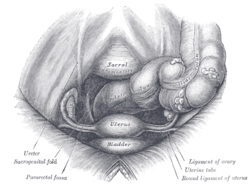
The rectouterine pouch is the extension of the peritoneum into the space between the posterior wall of the uterus and the rectum in the human female.

The posterior cricoarytenoid muscle is a intrinsic muscle of the larynx. It arises from the cricoid cartilage; it inserts onto the arytenoid cartilage of the same side. It is innervated by the recurrent laryngeal nerve. Each acts to open the vocal folds by pulling the vocal fold of the same side laterally. It participates in the production of sounds.

The superior sagittal sinus, within the human head, is an unpaired area along the attached margin of the falx cerebri. It allows blood to drain from the lateral aspects of anterior cerebral hemispheres to the confluence of sinuses. Cerebrospinal fluid drains through arachnoid granulations into the superior sagittal sinus and is returned to venous circulation.

The thyroarytenoid muscle is a broad, thin muscle that forms the body of the vocal fold and that supports the wall of the ventricle and its appendix. It functions to shorten the vocal folds.

The palmar aponeurosis invests the muscles of the palm, and consists of central, lateral, and medial portions.

The interphalangeal joints of the hand are the hinge joints between the phalanges of the fingers that provide flexion towards the palm of the hand.

In the human larynx, the cuneiform cartilages are two small, elongated pieces of yellow elastic cartilage, placed one on either side, in the aryepiglottic fold.

The anterior or lingual surface of the epiglottis is curved forward, and covered on its upper, free part by mucous membrane which is reflected on to the sides and root of the tongue, forming a median and two lateral glossoepiglottic folds; the lateral folds are partly attached to the wall of the pharynx.

The lateral umbilical fold is an elevation of the peritoneum lining the inner/posterior surface of the lower anterior abdominal wall formed by the underlying inferior epigastric artery and inferior epigastric vein which the peritoneum covers. Superiorly, the lateral umbilical fold ends where the vessels reach and enter the rectus sheath at the arcuate line of rectus sheath; in spite of the name, the lateral umbilical folds do not extend as far superiorly as the umbilicus. Inferiorly, it extends to just medial to the deep inguinal ring.

The ligament of the head of the femur is a weak ligament located in the hip joint. It is triangular in shape and somewhat flattened. The ligament is implanted by its apex into the anterosuperior part of the fovea capitis femoris and its base is attached by two bands, one into either side of the acetabular notch, and between these bony attachments it blends with the transverse ligament.

The fimbriated fold of tongue, also plica fimbriata, is a slight fold of the mucous membrane on the underside of the tongue which runs laterally on either side of the frenulum. The free edge of the fimbriated fold occasionally exhibits a series of fringe-like processes..

The medial umbilical ligament, cord of umbilical artery, or obliterated umbilical artery is a paired structure found in human anatomy. It is on the deep surface of the anterior abdominal wall, and is covered by the medial umbilical folds. It is different from the median umbilical ligament, a structure that represents the remnant of the embryonic urachus.

The medial umbilical fold is an elevation of the peritoneum lining the inner surface of the lower anterior abdominal wall formed by the underlying medial umbilical ligament which the peritoneum covers. Superiorly, the two medial umbilical folds converge towards the midline to meet and terminate at the umbilicus.
The pararectal fossa is an inferior-ward extension of the peritoneum on either side of the rectum. It is formed by a (sacrogenital) fold of peritoneum extending inferior-ward from the posterolateral pelvic wall. It represents a lateral extension of the rectouterine pouch in the female, and of rectovesical pouch in the male. It varies in size with the distension of the rectum.
In human female anatomy, the vesicouterine pouch, also uterovesicle pouch, is a fold of peritoneum over the uterus and the bladder. Like the rectouterine pouch, it is a female pelvic recess, but shallower and closer to the anterior fornix of the vagina.
The peritoneum of the anterior pelvic wall covers the superior surface of the bladder, and on either side of this viscus forms a depression, termed the paravesical fossa, which is limited laterally by the fold of peritoneum covering the ductus deferens.

The uterosacral ligaments are major ligaments of uterus that extend posterior-ward from the cervix to attach onto the sacrum.

The laryngeal ventricle, is a fusiform fossa, situated between the vestibular and vocal folds on either side, and extending nearly their entire length. There is also a sinus of Morgagni in the pharynx.

This article describes anatomical terminology that is used to describe the central and peripheral nervous systems - including the brain, brainstem, spinal cord, and nerves.