Articles related to anatomy include:

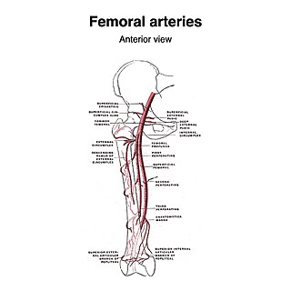
The femoral artery is a large artery in the thigh and the main arterial supply to the thigh and leg. The femoral artery gives off the deep femoral artery and descends along the anteromedial part of the thigh in the femoral triangle. It enters and passes through the adductor canal, and becomes the popliteal artery as it passes through the adductor hiatus in the adductor magnus near the junction of the middle and distal thirds of the thigh.

In human anatomy, the common iliac veins are formed by the external iliac veins and internal iliac veins. The left and right common iliac veins come together in the abdomen at the level of the fifth lumbar vertebra, forming the inferior vena cava. They drain blood from the pelvis and lower limbs.
The external iliac veins are large veins that connect the femoral veins to the common iliac veins. Their origin is at the inferior margin of the inguinal ligaments and they terminate when they join the internal iliac veins.

The internal iliac artery is the main artery of the pelvis.

The lateral cutaneous nerve of the thigh is a cutaneous nerve of the thigh. It originates from the dorsal divisions of the second and third lumbar nerves from the lumbar plexus. It passes under the inguinal ligament to reach the thigh. It supplies sensation to the skin on the lateral part of the thigh by an anterior branch and a posterior branch.

The iliolumbar artery is the first branch of the posterior trunk of the internal iliac artery.

The obturator artery is a branch of the internal iliac artery that passes antero-inferiorly on the lateral wall of the pelvis, to the upper part of the obturator foramen, and, escaping from the pelvic cavity through the obturator canal, it divides into an anterior branch and a posterior branch.

The median sacral artery is a small artery that arises posterior to the abdominal aorta and superior to its bifurcation.

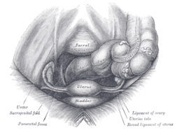
The ovarian artery is an artery that supplies oxygenated blood to the ovary in females. It arises from the abdominal aorta below the renal artery. It can be found within the suspensory ligament of the ovary, anterior to the ovarian vein and ureter.

The internal iliac vein begins near the upper part of the greater sciatic foramen, passes upward behind and slightly medial to the internal iliac artery and, at the brim of the pelvis, joins with the external iliac vein to form the common iliac vein.
The testicular artery is a branch of the abdominal aorta that supplies blood to the testis. It is a paired artery, with one for each of the testes.

The medial umbilical ligament is a paired structure found in human anatomy. It is on the deep surface of the anterior abdominal wall, and is covered by the medial umbilical folds. It is different from the median umbilical ligament, a structure that represents the remnant of the embryonic urachus.

The deep circumflex iliac artery is an artery in the pelvis that travels along the iliac crest of the pelvic bone.
The superficial iliac circumflex artery, the smallest of the cutaneous branches of the femoral artery, arises close to the superficial epigastric artery, and, piercing the fascia lata, runs lateralward, parallel with the inguinal ligament, as far as the crest of the ilium.

The superficial external pudendal artery is one of the three pudendal arteries. It arises from the medial side of the femoral artery, close to the superficial epigastric artery and superficial iliac circumflex artery.
The lateral sacral veins accompany the lateral sacral arteries on the anterior surface of the sacrum. They drain into the internal iliac vein. They communicate with each other via the sacral venous plexus.

In anatomy, the saphenous opening is an oval opening in the upper mid part of the fascia lata of the thigh. It lies 3–4 cm below and lateral to the pubic tubercle and is about 3 cm long and 1.5 cm wide.

The uterine vein is a vein of the uterus. It is found in the cardinal ligament. It drains into the internal iliac vein. It follows a similar course to the uterine artery. It helps to drain blood from the uterus, and removes waste from blood in the placenta during pregnancy.

The following outline is provided as an overview of and topical guide to human anatomy: