| Retroperitoneal space | |
|---|---|
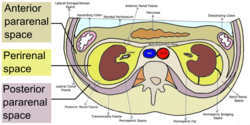 Horizontal plane through the kidneys, showing subdivisions of the retroperitoneal space. The anterior and posterior pararenal spaces have been exaggerated to provide representation of their relation to other retroperitoneal structures. | |
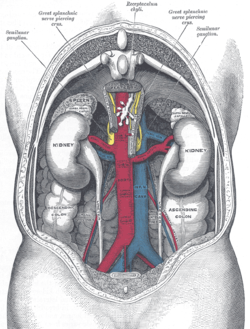 Human kidneys viewed from behind with spine removed | |
| Details | |
| Identifiers | |
| Latin | spatium retroperitoneale |
| MeSH | D012187 |
| TA98 | A10.1.01.002 |
| TA2 | 3814 |
| FMA | 15080 |
| Anatomical terminology | |
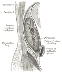
The retroperitoneal space (retroperitoneum) is the anatomical space (sometimes a potential space) behind (retro) the peritoneum. It has no specific delineating anatomical structures. Organs are retroperitoneal if they have peritoneum on their anterior side only. Structures that are not suspended by mesentery in the abdominal cavity and that lie between the parietal peritoneum and abdominal wall are classified as retroperitoneal. [1]
Contents
This is different from organs that are not retroperitoneal, which have peritoneum on their posterior side and are suspended by mesentery in the abdominal cavity.
The retroperitoneum can be further subdivided into the following: [2]
- Perirenal (or perinephric) space
- Anterior pararenal (or paranephric) space
- Posterior pararenal (or paranephric) space