
The spleen is an organ found in almost all vertebrates. Similar in structure to a large lymph node, it acts primarily as a blood filter. The word spleen comes from Ancient Greek σπλήν (splḗn).

In human anatomy, the mesentery, an organ that attaches the intestines to the posterior abdominal wall, comprises the double fold of the peritoneum. It helps in storing fat and allowing blood vessels, lymphatics, and nerves to supply the intestines.
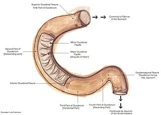
The suspensory muscle of duodenum is a thin muscle connecting the junction between the duodenum and jejunum, as well as the duodenojejunal flexure to connective tissue surrounding the superior mesenteric and coeliac arteries. The suspensory muscle most often connects to both the third and fourth parts of the duodenum, as well as the duodenojejunal flexure, although the attachment is quite variable.

The lesser omentum is the double layer of peritoneum that extends from the liver to the lesser curvature of the stomach, and to the first part of the duodenum. The lesser omentum is usually divided into these two connecting parts: the hepatogastric ligament, and the hepatoduodenal ligament.
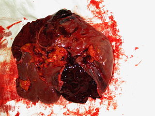
A splenic injury, which includes a ruptured spleen, is any injury to the spleen. The rupture of a normal spleen can be caused by trauma, such as a traffic collision.

The hepatic artery proper is the artery that supplies the liver and gallbladder. It raises from the common hepatic artery, a branch of the celiac artery.

The cystic artery is (usually) a branch of the right hepatic artery that provides arterial supply to the gallbladder and contributes arterial supply to the extrahepatic bile ducts.

In the anatomy of the human digestive tract, there are two colic flexures, or curvatures in the transverse colon. The right colic flexure is also known as the hepatic flexure, and the left colic flexure is also known as the splenic flexure. Note that "right" refers to the patient's anatomical right, which may be depicted on the left of a diagram.

The greater omentum is a large apron-like fold of visceral peritoneum that hangs down from the stomach. It extends from the greater curvature of the stomach, passing in front of the small intestines and doubles back to ascend to the transverse colon before reaching to the posterior abdominal wall. The greater omentum is larger than the lesser omentum, which hangs down from the liver to the lesser curvature. The common anatomical term "epiploic" derives from "epiploon", from the Greek epipleein, meaning to float or sail on, since the greater omentum appears to float on the surface of the intestines. It is the first structure observed when the abdominal cavity is opened anteriorly.

The duodenojejunal flexure or duodenojejunal junction, also known as the angle of Treitz, is the border between the duodenum and the jejunum.

The round ligament of the liver, ligamentum teres or ligamentum teres hepatis is a ligament that forms part of the free edge of the falciform ligament of the liver. It connects the liver to the umbilicus. It is the remnant of the left umbilical vein. The round ligament divides the left part of the liver into medial and lateral sections.

A trocar is a medical or veterinary device used in minimally invasive surgery. Trocars are typically made up of an awl, a cannula and often a seal. Some trocars also include a valve mechanism to allow for insufflation. Trocars are designed for placement through the chest and abdominal walls during thoracoscopic and laparoscopic surgery, and each trocar functions as a portal for the subsequent insertion of other endoscopic instruments such as grasper, scissors, stapler, electrocautery, suction tip, etc. — hence the more commonly used colloquial jargon "port". Trocars also allow passive evacuation of excess gas or fluid from organs within the body.
Hemoperitoneum is the presence of blood in the peritoneal cavity. The blood accumulates in the space between the inner lining of the abdominal wall and the internal abdominal organs. Hemoperitoneum is generally classified as a surgical emergency; in most cases, urgent laparotomy is needed to identify and control the source of the bleeding. In selected cases, careful observation may be permissible. The abdominal cavity is highly distensible and may easily hold greater than five liters of blood, or more than the entire circulating blood volume for an average-sized individual. Therefore, large-scale or rapid blood loss into the abdomen will reliably induce hemorrhagic shock and, if untreated, may rapidly lead to death.

In human anatomy, the median umbilical ligament is an unpaired midline ligamentous structure upon the lower inner surface of the anterior abdominal wall. It is covered by the median umbilical fold.
The paracolic gutters are peritoneal recesses – spaces between the colon and the abdominal wall.

The gastrosplenic ligament is part of the greater omentum extending between the stomach and the spleen. It contains several blood vessels.

An accessory spleen is a small nodule of splenic tissue found apart from the main body of the spleen. Accessory spleens are found in approximately 10 percent of the population and are typically around 1 centimetre in diameter. They may resemble a lymph node or a small spleen. They form either by the result of developmental anomalies or trauma. They are medically significant in that they may result in interpretation errors in diagnostic imaging or continued symptoms after therapeutic splenectomy. Polysplenia is the presence of multiple accessory spleens rather than one normal spleen.

Wandering spleen is a rare medical disease caused by the loss or weakening of the ligaments that help to hold the spleen stationary.

Peritoneal recesses are the spaces formed by peritoneum draping over viscera.
An exploratory laparotomy is a general surgical operation where the abdomen is opened and the abdominal organs are examined for injury or disease. It is the standard of care in various blunt and penetrating trauma situations in which there may be life-threatening internal injuries. It is also used in certain diagnostic situations, in which the operation is undertaken in search of a unifying cause for multiple signs and symptoms of disease, and in the staging of some cancers.

