The peritoneum is the serous membrane forming the lining of the abdominal cavity or coelom in amniotes and some invertebrates, such as annelids. It covers most of the intra-abdominal organs, and is composed of a layer of mesothelium supported by a thin layer of connective tissue. This peritoneal lining of the cavity supports many of the abdominal organs and serves as a conduit for their blood vessels, lymphatic vessels, and nerves.

The abdominal cavity is a large body cavity in humans and many other animals that contain organs. It is a part of the abdominopelvic cavity. It is located below the thoracic cavity, and above the pelvic cavity. Its dome-shaped roof is the thoracic diaphragm, a thin sheet of muscle under the lungs, and its floor is the pelvic inlet, opening into the pelvis.

The mesentery is an organ that attaches the intestines to the posterior abdominal wall and is formed by the double fold of peritoneum. It helps in storing fat and allowing blood vessels, lymphatics, and nerves to supply the intestines, among other functions.
The peritoneal cavity is a potential space between the parietal peritoneum and visceral peritoneum. The parietal and visceral peritonea are layers of the peritoneum named depending on their function/location. It is one of the spaces derived from the coelomic cavity of the embryo, the others being the pleural cavities around the lungs and the pericardial cavity around the heart.

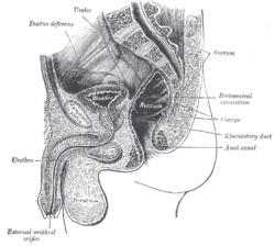
The rectouterine pouch is the extension of the peritoneum into the space between the posterior wall of the uterus and the rectum in the human female.

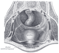
The internal obturator muscle or obturator internus muscle originates on the medial surface of the obturator membrane, the ischium near the membrane, and the rim of the pubis.

The common iliac artery is a large artery of the abdomen paired on each side. It originates from the aortic bifurcation at the level of the 4th lumbar vertebra. It ends in front of the sacroiliac joint, one on either side, and each bifurcates into the external and internal iliac arteries.

The iliopsoas muscle refers to the joined psoas major and the iliacus muscles. The two muscles are separate in the abdomen, but usually merge in the thigh. They are usually given the common name iliopsoas. The iliopsoas muscle joins to the femur at the lesser trochanter. It acts as the strongest flexor of the hip.

The abdomen is the part of the body between the thorax (chest) and pelvis, in humans and in other vertebrates. The abdomen is the front part of the abdominal segment of the torso. The area occupied by the abdomen is called the abdominal cavity. In arthropods, it is the posterior tagma of the body; it follows the thorax or cephalothorax.

The right colic artery is an artery of the abdomen, a branch of the superior mesenteric artery supplying the ascending colon. It divides into two terminal branches - an ascending branch and a descending branch - which form anastomoses with the middle colic artery, and ileocolic artery (respectively).

The greater omentum is a large apron-like fold of visceral peritoneum that hangs down from the stomach. It extends from the greater curvature of the stomach, passing in front of the small intestines and doubles back to ascend to the transverse colon before reaching to the posterior abdominal wall. The greater omentum is larger than the lesser omentum, which hangs down from the liver to the lesser curvature. The common anatomical term "epiploic" derives from "epiploon", from the Greek epipleein, meaning to float or sail on, since the greater omentum appears to float on the surface of the intestines. It is the first structure observed when the abdominal cavity is opened anteriorly.

The duodenojejunal flexure or duodenojejunal junction, also known as the angle of Treitz, is the border between the duodenum and the jejunum.

The pelvic cavity is a body cavity that is bounded by the bones of the pelvis. Its oblique roof is the pelvic inlet. Its lower boundary is the pelvic floor.

The pelvic brim is the edge of the pelvic inlet. It is an approximately Mickey Mouse head-shaped line passing through the prominence of the sacrum, the arcuate and pectineal lines, and the upper margin of the pubic symphysis.

The pubic tubercle is a prominent tubercle on the superior ramus of the pubis bone of the pelvis.

The superior rectal artery is an artery that descends into the pelvis to supply blood to the rectum.

The perimetrium is the outer serosal layer of the uterus, derived from the peritoneum overlying the uterine fundus, and can be considered a visceral peritoneum. It consists of a superficial layer of mesothelium, and a thin layer of loose connective tissue beneath it.
The paracolic gutters are peritoneal recesses – spaces between the colon and the abdominal wall.
In human female anatomy, the vesicouterine pouch, also uterovesicle pouch, is a fold of peritoneum over the uterus and the bladder. Like the rectouterine pouch, it is a female pelvic recess, but shallower and closer to the anterior fornix of the vagina.
In medicine, excavation has two meanings: