Seminal vesicles

Last updated

Seminal vesicle
Male anatomy 1.png
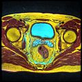
Cross-section of the lower abdomen in a male, showing parts of the urinary tract and male reproductive system, with the seminal vesicles seen top right
Gray1160.png
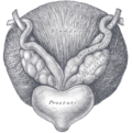
The seminal vesicles seen near the prostate, viewed from in front and above.
Details
Precursor Wolffian duct
System Male reproductive system
Artery Inferior vesical artery, middle rectal artery
Lymph External iliac lymph nodes, internal iliac lymph nodes
Identifiers
Latin vesiculae seminales, glandulae vesiculosae
MeSH D012669
TA98 A09.3.06.001
TA2 3631
FMA 19386
Anatomical terminology

The seminal vesicles (also called vesicular glands [1] or seminal glands) are a pair of convoluted tubular accessory glands that lie behind the urinary bladder of male mammals. They secrete fluid that partly composes the semen.

Contents

The vesicles are 5–10 cm in size, 3–5 cm in diameter, and are located between the bladder and the rectum. They have multiple outpouchings, which contain secretory glands, which join together with the vasa deferentia at the ejaculatory ducts. They receive blood from the vesiculodeferential artery, and drain into the vesiculodeferential veins. The glands are lined with column-shaped and cuboidal cells. The vesicles are present in many groups of mammals, but not marsupials, monotremes or carnivores.

Inflammation of the seminal vesicles is called seminal vesiculitis and most often is due to bacterial infection as a result of a sexually transmitted infection or following a surgical procedure. Seminal vesiculitis can cause pain in the lower abdomen, scrotum, penis or peritoneum, painful ejaculation, and blood in the semen. It is usually treated with antibiotics, although may require surgical drainage in complicated cases. Other conditions may affect the vesicles, including congenital abnormalities such as failure or incomplete formation, and, uncommonly, tumours.

The seminal vesicles have been described as early as the second century AD by Galen, although the vesicles only received their name much later, as they were initially described using the term from which the word prostate is derived.

Structure

The seminal vesicles are a pair of glands in males that are positioned below the urinary bladder and at the end of the vasa deferentia, where they enter the prostate. Each vesicle is a coiled and folded tube, with occasional outpouchings termed diverticula in its wall. [2] The lower part of the tube ends as a straight tube called the excretory duct, which joins with the vas deferens of that side of the body to form an ejaculatory duct. The ejaculatory ducts pass through the prostate gland before opening separately into the verumontanum of the prostatic urethra. [2] The vesicles are between 5–10 cm in size, 3–5 cm in diameter, and have a volume of around 13 mL. [3]

The vesicles receive blood supply from the vesiculodeferential artery, and also from the inferior vesical artery. The vesiculodeferential artery arises from the umbilical arteries, which branch directly from the internal iliac arteries. [3] Blood is drained into the vesiculodeferential veins and the inferior vesical plexus, which drain into the internal iliac veins. [3] Lymphatic drainage occurs along the venous routes, draining into the internal iliac nodes. [3]

The vesicles lie behind the bladder at the end of the vasa deferentia. They lie in the space between the bladder and the rectum; the bladder and prostate lie in front, the tip of the ureter as it enters the bladder above, and Denonvilliers' fascia and the rectum behind. [3]

Development

In the developing embryo, at the hind end lies a cloaca. This, over the fourth to the seventh week, divides into a urogenital sinus and the beginnings of the anal canal, with a wall forming between these two inpouchings called the urorectal septum. [4] Two ducts form next to each other that connect to the urogenital sinus; the mesonephric duct and the paramesonephric duct, which go on to form the reproductive tracts of the male and female respectively. [4]

In the male, under the influence of testosterone, the mesonephric duct proliferates, forming the epididymis, ductus deferens and, via a small outpouching near the developing prostate, the seminal vesicles. [4] Sertoli cells secrete anti-Müllerian hormone, which causes the paramesonephric duct to regress. [4]

The development and maintenance of the seminal vesicles, as well as their secretion and size/weight, are highly dependent on androgens. [5] [6] The seminal vesicles contain 5α-reductase, which metabolizes testosterone into its much more potent metabolite, dihydrotestosterone (DHT). [6] The seminal vesicles have also been found to contain luteinizing hormone receptors, and hence may also be regulated by the ligand of this receptor, luteinizing hormone. [6]

Microanatomy

The inner lining of the seminal vesicles (the epithelium) is made of a lining of interspersed column-shaped and cube-shaped cells. [7] There are varying descriptions of the lining as being pseudostratified and consisting of column-shaped cells only. [8] When viewed under a microscope, the cells are seen to have large bubbles in their interior. This is because their interior, called cytoplasm, contains lipid droplets involved in secretion during ejaculation. [7] The tissue of the seminal vesicles is full of glands, spaced irregularly. [7] As well as glands, the seminal vesicles contain smooth muscle and connective tissue. [7] This fibrous and muscular tissue surrounds the glands, helping to expel their contents. [3] The outer surface of the glands is covered in peritoneum. [3]

Function

The seminal vesicles secrete a significant proportion of the fluid that ultimately becomes semen. [9] Fluid is secreted from the ejaculatory ducts of the vesicles into the vas deferens, where it becomes part of semen. This then passes through the urethra, where it is ejaculated during a male sexual response. [8]

About 70-85% of the seminal fluid in humans originates from the seminal vesicles. [10] The fluid consists of nutrients including fructose and citric acid, prostaglandins, and fibrinogen. [9] Fructose is not produced anywhere else in the body except in the seminal vesicles. It provides a forensic test in rape cases.

Nutrients help support sperm until fertilisation occurs; prostaglandins may also assist by softening mucous of the cervix, and by causing reverse contractions of parts of the female reproductive tract such as the fallopian tubes, to ensure that sperm are less likely to be expelled. [9]

Clinical significance

Disease

Diseases of the seminal vesicles as opposed to that of prostate gland are extremely rare and are infrequently reported in the medical literature. [11]

Congenital anomalies associated with the seminal vesicles include failure to develop, either completely (agenesis) or partially (hypoplasia), and cysts. [12] [13] Failure of the vesicles to form is often associated with absent vas deferens, or an abnormal connection between the vas deferens and the ureter. [3] The seminal vesicles may also be affected by cysts, amyloidosis, and stones. [12] [13] Stones or cysts that become infected, or obstruct the vas deferens or seminal vesicles, may require surgical intervention. [8]

Seminal vesiculitis (also known as spermatocystitis) is an inflammation of the seminal vesicles, most often caused by bacterial infection. [14] Symptoms can include vague back or lower abdominal pain; pain of the penis, scrotum or peritoneum; painful ejaculation; blood in the semen on ejaculation; irritative and obstructive voiding symptoms; and impotence. [15] Infection may be due to sexually transmitted infections, as a complication of a procedure such as prostate biopsy. [8] It is usually treated with antibiotics. If a person experiences ongoing discomfort, transurethral seminal vesiculoscopy may be considered. [16] [17] Intervention in the form of drainage through the skin or surgery may also be required if the infection becomes an abscess. [8] The seminal vesicles may also be affected by tuberculosis, schistosomiasis and hydatid disease. [12] [13] These diseases are investigated, diagnosed and treated according to the underlying disease. [8]

Benign tumours of the seminal vesicles are rare. [8] When they do occur, they are usually papillary adenomas and cystadenomas. They do not cause elevation of tumour markers and are usually diagnosed based examination of tissue that has been removed after surgery. [8] Primary adenocarcinoma of the seminal vesicles, although rare, constitutes the most common malignant cancer of the seminal vesicles; [18] that said, the majority of malignant cancers affecting the vesicles are lesions that have extended into the vesicles from nearby parts of the body. [8] When adenocarcinoma occurs, it can cause blood in the urine, blood in the semen, painful urination, urinary retention, or even urinary obstruction. [8] Adenocarcinomata are usually diagnosed after they are excised, based on tissue diagnosis. [8] Some produce the tumour marker Ca-125, which can be used to monitor for reoccurence afterwards. [8] Even rarer neoplasms include sarcoma, squamous cell carcinoma, yolk sac tumor, neuroendocrine carcinoma, paraganglioma, epithelial stromal tumors and lymphoma. [18]

Investigations

Symptoms due to diseases of the seminal vesicles may be vague and not able to be specifically attributable to the vesicles themselves; additionally, some conditions such as tumours or cysts may not cause any symptoms at all. [8] When diseases is suspected, such as due to pain on ejaculation, blood in the urine, infertility, due to urinary tract obstruction, further investigations may be conducted. [8]

A digital rectal examination, which involves a finger inserted by a medical practitioner through the anus, may cause greater than usual tenderness of the prostate gland, or may reveal a large seminal vesicle. [8] Palpation is dependent on the length of index finger as seminal vesicles are located above the prostate gland and retrovesical (behind the bladder).

A urine specimen may be collected, and is likely to demonstrate blood within the urine. [8] Laboratory examination of seminal vesicle fluid requires a semen sample, e.g. for semen culture or semen analysis. Fructose levels provide a measure of seminal vesicle function and, if absent, bilateral agenesis or obstruction is suspected. [12]

Imaging of the vesicles is provided by medical imaging; either by transrectal ultrasound, CT or MRI scans. [8] An examination using cystoscopy, where a flexible tube is inserted in the urethra, may show disease of the vesicles because of changes in the normal appearance of the nearby bladder trigone, or prostatic urethra. [8]

Other animals

The evolution of seminal vesicles may have been influenced by sexual selection. They occur in many groups of mammals, [19] but are absent in marsupials, [20] monotremes, and carnivorans. [21] [22] The function is similar in all mammals they are present in, which is to secrete a fluid as part of semen that is ejaculated during the sexual response. [19]

History

The action of the seminal vesicles has been described as early the second century AD by Galen, as "glandular bodies" that secrete substances alongside semen during reproduction. [21] By the time of Herophilus the presence of the glands and associated ducts had been described. [21] Around the time of the early 17th century the word used to describe the vesicles, parastatai, eventually and unambiguously was used to refer to the prostate gland, rather than the vesicles. [21] The first time the prostate was portrayed in an individual drawing was by Reiner De Graaf in 1678. [21]

The first described use of laparoscopic surgery on the vesicles was described in 1993; this is now the preferred approach because of decreased pain, complications, and a shorter hospital stay. [8]

Additional images

See also

Related Research Articles

<span class="mw-page-title-main">Urethra</span> Tube that connects the urinary bladder to the external urethral orifice

The urethra is a tube that connects the mammalian urinary bladder to the urinary meatus. Male and female placental mammals release urine through the urethra during urination, but males also release semen through the urethra during ejaculation.

<span class="mw-page-title-main">Prostate</span> Gland of the male reproductive system in most mammals

The prostate is both an accessory gland of the male reproductive system and a muscle-driven mechanical switch between urination and ejaculation. It is found in all male mammals. It differs between species anatomically, chemically, and physiologically. Anatomically, the prostate is found below the bladder, with the urethra passing through it. It is described in gross anatomy as consisting of lobes and in microanatomy by zone. It is surrounded by an elastic, fibromuscular capsule and contains glandular tissue, as well as connective tissue.

<span class="mw-page-title-main">Skene's gland</span> Glands located on the anterior wall of the vagina

In female human anatomy, Skene's glands or the Skene glands are glands located around the lower end of the urethral meatus. The glands are surrounded by tissue that swells with blood during sexual arousal, and secrete a fluid from openings near the urethra, particularly during orgasm.

<span class="mw-page-title-main">Bulbourethral gland</span> Gland in males to help with sperm health

The bulbourethral glands or Cowper's glands are two small exocrine and accessory glands in the reproductive system of many male mammals. They are homologous to Bartholin's glands in females. The bulbourethral glands are responsible for producing a pre-ejaculate fluid called Cowper's fluid, which is secreted during sexual arousal, neutralizing the acidity of the urethra in preparation for the passage of sperm cells. The paired glands are found adjacent to the urethra just below the prostate, seen best by screening (medicine) MRI as a tool in preventative healthcare in males. Screening MRI may be performed when there is a positive prostate-specific antigen on basic laboratory tests. Prostate cancer is the second-most common cause of cancer-related mortality in males in the USA.

<span class="mw-page-title-main">Ureter</span> Tubes used in the urinary system in most animals

The ureters are tubes made of smooth muscle that propel urine from the kidneys to the urinary bladder. In a human adult, the ureters are usually 20–30 cm (8–12 in) long and around 3–4 mm (0.12–0.16 in) in diameter. The ureter is lined by urothelial cells, a type of transitional epithelium, and has an additional smooth muscle layer that assists with peristalsis in its lowest third.

<span class="mw-page-title-main">Retrograde ejaculation</span> Redirection of ejaculated semen into the urinary bladder

Retrograde ejaculation occurs when semen which would be ejaculated via the urethra is redirected to the urinary bladder. Normally, the sphincter of the bladder contracts before ejaculation, sealing the bladder which besides inhibiting the release of urine also prevents a reflux of seminal fluids into the male bladder during ejaculation. The semen is forced to exit via the urethra, the path of least resistance. When the bladder sphincter does not function properly, retrograde ejaculation may occur. It can also be induced deliberately by a male as a primitive form of male birth control or as part of certain alternative medicine practices. The retrograde-ejaculated semen, which goes into the bladder, is excreted with the next urination.

<span class="mw-page-title-main">Epididymis</span> Tube that connects a testicle to a vas deferens

The epididymis is an elongated tubular structure attached to the posterior side of each one of the two male reproductive glands, the testicles. It is a single, narrow, tightly coiled tube in adult humans, 6 to 7 centimetres in length; uncoiled the tube would be approximately 6 m long. It connects the testicle to the vas deferens in the male reproductive system. The epididymis serves as an interconnection between the multiple efferent ducts at the rear of a testicle (proximally), and the vas deferens (distally). Its primary function is the storage, maturation and transport of sperm cells.

<span class="mw-page-title-main">Vas deferens</span> Part of the male reproductive system of many vertebrates

The vas deferens, with the more modern name ductus deferens, is part of the male reproductive system of many vertebrates. The ducts transport sperm from the epididymides to the ejaculatory ducts in anticipation of ejaculation. The vas deferens is a partially coiled tube which exits the abdominal cavity through the inguinal canal.

<span class="mw-page-title-main">Ejaculatory duct</span> Male anatomical structures

The ejaculatory ducts are paired structures in the male reproductive system. Each ejaculatory duct is formed by the union of the vas deferens with the duct of the seminal vesicle. They pass through the prostate, and open into the urethra above the seminal colliculus. During ejaculation, semen passes through the prostate gland, enters the urethra and exits the body via the urinary meatus.

<span class="mw-page-title-main">Mesonephric duct</span> Paired organ in mammals

The mesonephric duct, also known as the Wolffian duct, archinephric duct, Leydig's duct or nephric duct, is a paired organ that develops in the early stages of embryonic development in humans and other mammals. It is an important structure that plays a critical role in the formation of male reproductive organs. The duct is named after Caspar Friedrich Wolff, a German physiologist and embryologist who first described it in 1759.

<span class="mw-page-title-main">Male reproductive system</span> Reproductive system of the human male

The male reproductive system consists of a number of sex organs that play a role in the process of human reproduction. These organs are located on the outside of the body, and within the pelvis.

<span class="mw-page-title-main">Human reproductive system</span> Organs involved in reproduction

The human reproductive system includes the male reproductive system which functions to produce and deposit sperm; and the female reproductive system which functions to produce egg cells, and to protect and nourish the fetus until birth. Humans have a high level of sexual differentiation. In addition to differences in nearly every reproductive organ, there are numerous differences in typical secondary sex characteristics.

Aspermia is the complete lack of semen with ejaculation. It is associated with infertility.

Hypospermia is a condition in which a man has an unusually low ejaculate volume, less than 1.5 mL. It is the opposite of hyperspermia, which is a semen volume of more than 5.5 mL. It should not be confused with oligospermia, which means low sperm count. Normal ejaculate when a man is not drained from prior sex and is suitably aroused is around 1.5–6 mL, although this varies greatly with mood, physical condition, and sexual activity. Of this, around 1% by volume is sperm cells. The U.S.-based National Institutes of Health defines hypospermia as a semen volume lower than 2 mL on at least two semen analyses.

<span class="mw-page-title-main">Seminal colliculus</span> Part of the human male reproductive organs

The seminal colliculus, or verumontanum, of the prostatic urethra is a landmark distal to the entrance of the ejaculatory ducts. Verumontanum is translated from Latin to mean 'mountain ridge', a reference to the distinctive median elevation of urothelium that characterizes the landmark on magnified views.

<span class="mw-page-title-main">Human penis</span> Human male external reproductive organ

In human anatomy, the penis is an external male sex organ that additionally serves as the urinary duct. The main parts are the root, body, the epithelium of the penis including the shaft skin, and the foreskin covering the glans. The body of the penis is made up of three columns of tissue: two corpora cavernosa on the dorsal side and corpus spongiosum between them on the ventral side. The human male urethra passes through the prostate gland, where it is joined by the ejaculatory duct, and then through the penis. The urethra traverses the corpus spongiosum, and its opening, the meatus, lies on the tip of the glans. It is a passage both for urination and ejaculation of semen.

The development of the reproductive system is the part of embryonic growth that results in the sex organs and contributes to sexual differentiation. Due to its large overlap with development of the urinary system, the two systems are typically described together as the genitourinary system.

<span class="mw-page-title-main">Semen</span> Reproductive biofluid of male or hermaphroditic animals

Semen, also known as seminal fluid, is an organic bodily fluid that contains spermatozoa. Spermatozoa are secreted by the male gonads and other sexual organs of male or hermaphroditic animals and can fertilize the female ovum. Semen is produced and originates from the seminal vesicle, which is located in the pelvis. The process that results in the discharge of semen from the urethral orifice is called ejaculation. In humans, seminal fluid contains several components besides spermatozoa: proteolytic and other enzymes as well as fructose are elements of seminal fluid which promote the survival of spermatozoa and provide a medium through which they can move or "swim". The fluid is adapted to be discharged deep into the vagina, so the spermatozoa can pass into the uterus and form a zygote with an egg.

Ejaculatory duct obstruction (EDO) is a pathological condition which is characterized by the obstruction of one or both ejaculatory ducts. Thus, the efflux of semen is not possible. It can be congenital or acquired. It is a cause of male infertility and/or pelvic pain. Ejaculatory duct obstruction must not be confused with an obstruction of the vas deferens.

<span class="mw-page-title-main">Male accessory gland</span> Sexual gland in males

Male accessory glands (MAG) in humans are the seminal vesicles, prostate gland, and the bulbourethral glands. In insects, male accessory glands produce products that mix with the sperm to protect and preserve them, including seminal fluid proteins. Some insecticides can induce an increase in the protein content of the male accessory glands of certain types of insects. This has the unintended effect of increasing the number of offspring they produce.

References

  1. Rowen D. Frandson; W. Lee Wilke; Anna Dee Fails (2009). "Anatomy of the Male Reproductive System". Anatomy and Physiology of Farm Animals (7th ed.). John Wiley and Sons. p. 409. ISBN   978-0-8138-1394-3.
  2. 1 2 Michael H. Ross; Wojciech Pawlina (2010). "Male Reproductive System". Histology: A Text and Atlas, with Correlated Cell and Molecular Biology (6th ed.). Wolters Kluwer/Lippincott Williams & Wilkins Health. p. 828. ISBN   978-0781772006.
  3. 1 2 3 4 5 6 7 8 Standring, Susan, ed. (2016). "Seminal vesicles". Gray's anatomy : the anatomical basis of clinical practice (41st ed.). Philadelphia. pp. 1279–1280. ISBN   9780702052309. OCLC   920806541.{{cite book}}: CS1 maint: location missing publisher (link)
  4. 1 2 3 4 Sadley, TW (2019). "Genital ducts". Langman's medical embryology (14th ed.). Philadelphia: Wolters Kluwer. pp. 271–5. ISBN   9781496383907.
  5. B. Fey; F. Heni; A. Kuntz; D. F. McDonald; L. Quenu; L. G. jr. Wesson; C. Wilson (6 December 2012). Physiologie und Pathologische Physiologie / Physiology and Pathological Physiology / Physiologie Normale et Pathologique. Springer Science & Business Media. pp. 611–. ISBN   978-3-642-46018-0.
  6. 1 2 3 Gonzales GF (2001). "Function of seminal vesicles and their role on male fertility". Asian J. Androl. 3 (4): 251–8. PMID   11753468.
  7. 1 2 3 4 Young, Barbara; O'Dowd, Geraldine; Woodford, Phillip (2013). "Male reproductive system". Wheater's functional histology: a text and colour atlas (6th ed.). Philadelphia: Elsevier. p. 346. ISBN   9780702047473.
  8. 1 2 3 4 5 6 7 8 9 10 11 12 13 14 15 16 17 18 19 Arthur D. Smith (Editor), Glenn Preminger (Editor), Gopal H. Badlani (Editor), Louis R. Kavoussi (Editor) (2019). "112. Laparoscopic and Robotic Surgery of the Seminal Vessels". Smith's textbook of endourology (4th ed.). John Wiley & Sons Ltd. pp. 1292–1298. ISBN   9781119245193.{{cite book}}: |last1= has generic name (help)CS1 maint: multiple names: authors list (link)
  9. 1 2 3 Hall, John E (2016). "Function of the seminal vesicles". Guyton and Hall textbook of medical physiology (13th ed.). Philadelphia: Elsevier. p. 1024. ISBN   978-1-4557-7016-8.
  10. Kierszenbaum, Abraham L.; Tres, Laura (2011). "Chapter 21: Sperm Transport and Maturation". Histology and Cell Biology: An Introduction to Pathology (3rd ed.). St. Louis [u.a.]: Mosby. p. 624. ISBN   978-0323078429.
  11. Dagur G, Warren K, Suh Y, Singh N, Khan SA. Detecting diseases of neglected seminal vesicles using imaging modalities: A review of current literature. Int J Reprod Biomed. 2016;14(5):293-302.
  12. 1 2 3 4 El-Hakim, Assaad (13 November 2006). "Diagnosis and Treatment of Disorders of the Ejaculatory Ducts and Seminal Vesicles". In Smith, Arthur D. (ed.). Smith's Textbook of Endourology (2nd ed.). Wiley-Blackwell. pp. 759–766. ISBN   978-1550093650.
  13. 1 2 3 "Seminal vesicle diseases". Geneva Foundation for Medical Education and Research. Archived from the original on 2014-04-26.
  14. "Is seminal vesiculitis a discrete disease entity? Clinical and microbiological study of seminal vesiculitis in patients with acute epididymitis". 4 July 2023.
  15. Zeitlin, S. I.; Bennett, C. J. (1 November 1999). "Chapter 25: Seminal vesiculitis". In Curtis Nickel, J. (ed.). Textbook of Prostatitis. CRC Press. pp. 219–225. ISBN   9781901865042.
  16. La Vignera S (October 2011). "Male accessory gland infection and sperm parameters". International Journal of Andrology. 34 (5pt2): e330–47. doi: 10.1111/j.1365-2605.2011.01200.x . PMID   21696400.
  17. Bianjiang Liu; Jie Li; Pengchao Li; Jiexiu Zhang; Ninghong Song; Zengjun Wang; Changjun Yin (February 2014). "Transurethral seminal vesiculoscopy in the diagnosis and treatment of intractable seminal vesiculitis". The Journal of International Medical Research. 42 (1): 236–42. doi: 10.1177/0300060513509472 . PMID   24391141.
  18. 1 2 Katafigiotis, Ioannis; Sfoungaristos, Stavros; Duvdevani, Mordechai; Mitsos, Panagiotis; Roumelioti, Eleni; Stravodimos, Konstantinos; Anastasiou, Ioannis; Constantinides, Constantinos A. (31 March 2016). "Primary adenocarcinoma of the seminal vesicles. A review of the literature" (PDF). Archivio Italiano di Urologia e Andrologia. 88 (1): 47–51. doi: 10.4081/aiua.2016.1.47 . ISSN   1124-3562. PMID   27072175.
  19. 1 2 Kardong, Kenneth (2019). "Reproductive system". Vertebrates : comparative anatomy, function, evolution (8th ed.). New York: McGraw-Hill. p. 564. ISBN   9781260092042.
  20. Tyndale-Biscoe, C. Hugh; Renfree, Marilyn (1987-01-30). Reproductive Physiology of Marsupials. Cambridge University Press. ISBN   978-0-521-33792-2.
  21. 1 2 3 4 5 Josef Marx, Franz; Karenberg, Axel (1 February 2009). "History of the Term Prostate". The Prostate. 69 (2): 208–213. doi:10.1002/pros.20871. PMID   18942121. S2CID   44922919. The humor produced in those glandular bodies is poured into the urinary passage in the male along with semen and its uses are to excite to the sexual act, to make coitus pleasurable, and to moisten the urinary passageway.
  22. Dixson, Alan F. "Sexual selection and evolution of the seminal vesicles in primates." Folia Primatologica 69.5 (1998): 300-306.