Fibrous joint

Last updated
Fibrous joint
904 Fibrous Joints.jpg
Fibrous joints
Details
Identifiers
Latin articulatio fibrosa, junctura fibrosa
TA98 A03.0.00.004
TA2 1517
FMA 7492
Anatomical terminology

In anatomy, fibrous joints are joints connected by fibrous tissue, consisting mainly of collagen. These are fixed joints where bones are united by a layer of white fibrous tissue of varying thickness. In the skull, the joints between the bones are called sutures. Such immovable joints are also referred to as synarthroses.

Contents

Types

Most fibrous joints are also called "fixed" or "immovable". These joints have no joint cavity and are connected via fibrous connective tissue.

Sutures

Side view of the skull Gray188.png
Side view of the skull
Human skull side sutures right Human skull side suturas right.svg
Human skull side sutures right

A suture is a type of fibrous joint that is only found in the skull (cranial suture). The bones are bound together by Sharpey's fibres. A tiny amount of movement is permitted at sutures, which contributes to the compliance and elasticity of the skull. These joints are synarthroses. [1] It is normal for many of the bones of the skull to remain unfused at birth. The fusion of the skull's bones before birth is known as craniosynostosis. The term "fontanelle" is used to describe the resulting "soft spots". The relative positions of the bones continue to change during the life of the adult (though less rapidly), which can provide useful information in forensics and archaeology. In old age, cranial sutures may ossify (turn to bone) completely. [3] The joints between the teeth and jaws (gomphoses) and the joint between the mandible and the cranium, the temporomandibular joint, form the only non-sutured joints in the skull.

Types of sutures

List of sutures

Cranial sutures viewed from top of head Sutures from top.png
Cranial sutures viewed from top of head

Most sutures are named for the bones they articulate, but some have special names of their own.

Visible from the side

Visible from the front or above

Visible from below or inside

Syndesmosis

A syndesmosis is a slightly mobile [4] fibrous joint in which bones such as the tibia and fibula are joined together by connective tissue. An example is the distal tibiofibular joint. Injuries to the ankle syndesmosis are commonly known as a "high ankle sprain". Although the syndesmosis is a joint, in the literature the term syndesmotic injury is used to describe injury of the syndesmotic ligaments. It comes from the Greek σύν, syn (meaning "with") and δεσμός, desmos (meaning "a band"). [5] Syndesmosis sprains have received increasing recognition during recent years because of a heightened awareness of the mechanism, symptoms, and signs of injury. [6]

Diagnosis of a syndesmotic injury

Diagnosis of syndesmosis injuries by physical examination is often straightforward. Physical examination findings that are often positive include the squeeze test and the external rotation test. Patients with high-grade syndesmosis injuries often cannot perform a single-leg heel raise. Patients report pain in varying degrees over the anterior and often posterior distal fibular joint. [7]

Syndesmotic tear

The severity of acute syndesmosis injury is rated from grade I to III by several authors. A grade I injury is a partial anteroinferior tibiofibular ligament tear, meaning the exorotation and squeeze tests are negative for this grade. Grade II injury is a complete anteroinferior tibiofibular ligament and inferior interosseous ligament tear, meaning that squeeze test and exorotation are positive. This results in the injury being stabilized with immobilization but not operatively stabilized. A grade III injury is a complete anteroinferior tibiofibular ligament tear including a (partial) interosseous ligament tear and deltoid ligament avulsion, meaning the joint is unstable and positive on the exorotation and squeeze tests. This grade requires operative stabilization. [8] If the syndesmosis is torn apart as result of bone fracture, surgeons will sometimes fix the relevant bones together with a syndesmotic screw, temporarily replacing the syndesmosis, or with a tightrope fixation, which is called syndesmosis procedure. [9] [10] The screw inhibits normal movement of the bones and, thereby, the corresponding joint(s). When the natural articulation is healed, the screw may be removed. The tightrope fixation with elastic fiberwire suture on the other hand allows physiologic motion of the ankle and may be permanent.

Gomphosis

The teeth, viewed from the right Gray1003.png
The teeth, viewed from the right

A gomphosis, also known as a dentoalveolar syndesmosis, [11] or 'peg and socket joint' [12] is a joint that binds the teeth to bony teeth sockets in the maxillary bone and mandible. The fibrous connection between a tooth and its socket is a periodontal ligament. Specifically, the connection is made between the maxilla or mandible to the cementum of the tooth.

The motion of a gomphosis is minimal, though considerable movement can be achieved over time—the basis of using braces to realign teeth. The joint can be considered a synarthrosis. [13]

The gomphosis is the only joint-type in which a bone does not join another bone, as teeth are not technically bone. In modern, more anatomical, joint classification, the gomphosis is simply considered a fibrous joint because the tissue linking the structures is ligamentous. It has been suggested that this permanent soft-tissue attachment was a critical requisite in the evolution of the mammalian (synapsid) tusk. [14]

A gomphosis is a specialized fibrous joint in which a conical process or peg of one bone fits into a hole or socket in another bone. (gomphos is a Greek word meaning bolt). Small quantity of fibrous tissue holds the bones together. No movement is possible at such peg-and-socket joints.

Related Research Articles

<span class="mw-page-title-main">Skull</span> Bony structure that forms the head in vertebrates

The skull is a bone protective cavity for the brain. The skull is composed of four types of bone i.e., cranial bones, facial bones, ear ossicles and hyoid bone, however two parts are more prominent: the cranium and the mandible. In humans, these two parts are the neurocranium (braincase) and the viscerocranium that includes the mandible as its largest bone. The skull forms the anterior-most portion of the skeleton and is a product of cephalisation—housing the brain, and several sensory structures such as the eyes, ears, nose, and mouth. In humans, these sensory structures are part of the facial skeleton.

<span class="mw-page-title-main">Fontanelle</span> Anatomical feature of the infant human skull

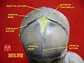
A fontanelle is an anatomical feature of the infant human skull comprising soft membranous gaps (sutures) between the cranial bones that make up the calvaria of a fetus or an infant. Fontanelles allow for stretching and deformation of the neurocranium both during birth and later as the brain expands faster than the surrounding bone can grow. Premature complete ossification of the sutures is called craniosynostosis.

Articles related to anatomy include:

<span class="mw-page-title-main">Tibia</span> Leg bone in vertebrates

The tibia, also known as the shinbone or shankbone, is the larger, stronger, and anterior (frontal) of the two bones in the leg below the knee in vertebrates ; it connects the knee with the ankle. The tibia is found on the medial side of the leg next to the fibula and closer to the median plane. The tibia is connected to the fibula by the interosseous membrane of leg, forming a type of fibrous joint called a syndesmosis with very little movement. The tibia is named for the flute tibia. It is the second largest bone in the human body, after the femur. The leg bones are the strongest long bones as they support the rest of the body.

<span class="mw-page-title-main">Occipital bone</span> Skull bone, lower back of cranium

The occipital bone is a cranial dermal bone and the main bone of the occiput. It is trapezoidal in shape and curved on itself like a shallow dish. The occipital bone overlies the occipital lobes of the cerebrum. At the base of the skull in the occipital bone, there is a large oval opening called the foramen magnum, which allows the passage of the spinal cord.

<span class="mw-page-title-main">Parietal bone</span> Bone in the human skull which, when joined together, forms the sides and roof of the cranium

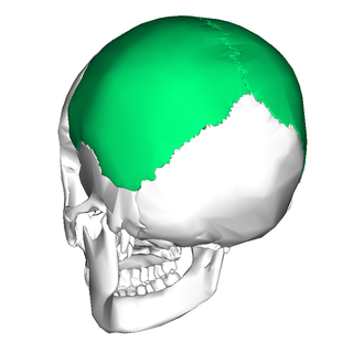
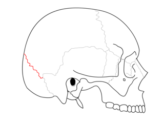
The parietal bones are two bones in the skull which, when joined at a fibrous joint, form the sides and roof of the cranium. In humans, each bone is roughly quadrilateral in form, and has two surfaces, four borders, and four angles. It is named from the Latin paries (-ietis), wall.

<span class="mw-page-title-main">Fibula</span> Leg bone in vertebrates

The fibula or calf bone is a leg bone on the lateral side of the tibia, to which it is connected above and below. It is the smaller of the two bones and, in proportion to its length, the most slender of all the long bones. Its upper extremity is small, placed toward the back of the head of the tibia, below the knee joint and excluded from the formation of this joint. Its lower extremity inclines a little forward, so as to be on a plane anterior to that of the upper end; it projects below the tibia and forms the lateral part of the ankle joint.

<span class="mw-page-title-main">Maisonneuve fracture</span> Medical condition

The Maisonneuve fracture is a spiral fracture of the proximal third of the fibula associated with a tear of the distal tibiofibular syndesmosis and the interosseous membrane. There is an associated fracture of the medial malleolus or rupture of the deep deltoid ligament of the ankle. This type of injury can be difficult to detect.

<span class="mw-page-title-main">Syndesmosis</span> Type of fibrous joint

A syndesmosis is a type of fibrous joint in which two parallel bones are united to each other by fibrous connective tissue. The gap between the bones may be narrow, with the bones joined by ligaments, or the gap may be wide and filled in by a broad sheet of connective tissue called an interosseous membrane. The syndesmoses found in the forearm and leg serve to unite parallel bones and prevent their separation.

<span class="mw-page-title-main">Synarthrosis</span> Immobile joint type

A synarthrosis is a type of joint which allows no movement under normal conditions. Sutures and gomphoses are both synarthroses. Joints which allow more movement are called amphiarthroses or diarthroses. Syndesmoses are considered to be amphiarthrotic, because they allow a small amount of movement.

<span class="mw-page-title-main">Sagittal suture</span> Midline joint between the parietal bones of the skull

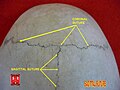
The sagittal suture, also known as the interparietal suture and the sutura interparietalis, is a dense, fibrous connective tissue joint between the two parietal bones of the skull. The term is derived from the Latin word sagitta, meaning arrow.

<span class="mw-page-title-main">Lambdoid suture</span> Connective tissue between the parietal bones and the occipital bone of the skull

The lambdoid suture is a dense, fibrous connective tissue joint on the posterior aspect of the skull that connects the parietal bones with the occipital bone. It is continuous with the occipitomastoid suture.

<span class="mw-page-title-main">Coronal suture</span> Connective tissue between the parietal bones and the frontal bone of the skull

The coronal suture is a dense, fibrous connective tissue joint that separates the two parietal bones from the frontal bone of the skull.

<span class="mw-page-title-main">Ankle fracture</span> Medical condition

An ankle fracture is a break of one or more of the bones that make up the ankle joint. Symptoms may include pain, swelling, bruising, and an inability to walk on the injured leg. Complications may include an associated high ankle sprain, compartment syndrome, stiffness, malunion, and post-traumatic arthritis.

<span class="mw-page-title-main">Anterior tibiofibular ligament</span>

The anterior ligament of the lateral malleolus is a flat, trapezoidal band of fibers, broader below than above, which extends obliquely downward and lateralward between the adjacent margins of the tibia and fibula, on the front aspect of the syndesmosis.

<span class="mw-page-title-main">Interosseous membrane of leg</span>

The interosseous membrane of the leg extends between the interosseous crests of the tibia and fibula, helps stabilize the Tib-Fib relationship and separates the muscles on the front from those on the back of the leg.

<span class="mw-page-title-main">Calvaria (skull)</span> Top part of the skull

The calvaria is the top part of the skull. It is the superior part of the neurocranium and covers the cranial cavity containing the brain. It forms the main component of the skull roof.

In arthropod and vertebrate anatomy, the vertex is the highest point of the head.

<span class="mw-page-title-main">Outline of human anatomy</span> Overview of and topical guide to human anatomy

The following outline is provided as an overview of and topical guide to human anatomy:

The fetal head, from an obstetrical viewpoint, and in particular its size, is important because an essential feature of labor is the adaptation between the fetal head and the maternal bony pelvis. Only a comparatively small part of the head at term is represented by the face. The rest of the head is composed of the firm skull, which is made up of two frontal, two parietal, and two temporal bones, along with the upper portion of the occipital bone and the wings of the sphenoid.

References

  1. 1 2 "Module – Introduction to Joints". Archived from the original on 2007-12-17. Retrieved 2008-01-29.
  2. Tomco R. "Fibrous Joints". AnatomyOne. Amirsys, Inc. Archived from the original on 2013-01-16. Retrieved 2012-09-28.
  3. Harth S, Obert M, Ramsthaler F, Reuss C, Traupe H, Verhoff MA (April 2009). "Estimating age by assessing the ossification degree of cranial sutures with the aid of Flat-Panel-CT". Legal Medicine. 11 Suppl 1 (Supp.1): S186-9. doi:10.1016/j.legalmed.2009.01.091. PMID   19261532.
  4. Juneja, Pallavi; Munjal, Akul; Hubbard, John B. (2022), "Anatomy, Joints", StatPearls, Treasure Island (FL): StatPearls Publishing, PMID   29939670 , retrieved 2022-04-26
  5. Arnold MA, Bryce D. "Arnold's Glossary of Anatomy". The University of Sydney.
  6. Jones MH, Amendola A (February 2007). "Syndesmosis sprains of the ankle: a systematic review". Clinical Orthopaedics and Related Research. 455: 173–5. doi:10.1097/BLO.0b013e31802eb471. PMID   17146360. S2CID   13598905.
  7. Metzler AV, Johnson DL (March 2013). "Dynamically unstable syndesmosis injuries". Orthopedics. 36 (3): 209–11. doi:10.3928/01477447-20130222-07. PMID   23464940.
  8. Valkering KP, Vergroesen DA, Nolte PA (December 2012). "Isolated syndesmosis ankle injury". Orthopedics. 35 (12): e1705–10. doi:10.3928/01477447-20121120-13. PMID   23218625.
  9. Porucznik MA (May 2008). "Screw vs. tightrope fixation for syndesmotic fractures". AAOS Now. American Academy of Orthopaedic Surgeons. Archived from the original on 14 June 2010.
  10. "Arthrex - TightRope®". www.arthrex.com.
  11. "Dentoalveolar syndesmosis". TheFreeDictionary.com.
  12. Betts, J. Gordon (2013). "9.2 Fibrous joints". Anatomy & physiology. Houston, Texas: OpenStax. ISBN   978-1-947172-04-3 . Retrieved 16 April 2023.
  13. Billy A. "Articulations". Douglas College. Archived from the original on 2007-01-04. Retrieved 2008-01-29.
  14. Whitney, M.R.; Angielczyk, K.D.; Peecook, B.R.; Sidor, C.A. (2021). "The evolution of the synapsid tusk: insights from dicynodont therapsid tusk histology". Proceedings of the Royal Society B. 288 (1961): 20211670. doi:10.1098/rspb.2021.1670. PMC   8548784 . PMID   34702071.