Pisometacarpal ligament

Last updated
Pisometacarpal ligament
1121 Intrinsic Muscles of the Hand Deep LD.png
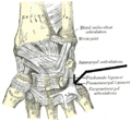
Deep muscles of the right hand, with pisometacarpal ligament at top right. Dorsal view.
Details
From Pisiform
To Fifth metacarpal
Identifiers
Latin ligamentum pisometacarpeum
TA98 A03.5.11.109
TA2 1826
FMA 42305
Anatomical terminology

The pisometacarpal ligament joins the pisiform to the base of the fifth metacarpal bone. It is a continuation of the tendon of the flexor carpi ulnaris.

Contents

Additional images

Related Research Articles

Flexor carpi ulnaris muscle

The flexor carpi ulnaris (FCU) is a muscle of the forearm that flexes and adducts the wrist joint.

Inguinal triangle

In human anatomy, the inguinal triangle is a region of the abdominal wall. It is also known by the eponym Hesselbach's triangle, after Franz Kaspar Hesselbach.

Round ligament of uterus

The round ligament of the uterus originates at the uterine horns, in the parametrium. The round ligament exits the pelvis via the deep inguinal ring, passes through the inguinal canal and continues on to the labia majora where its fibers spread and mix with the tissue of the mons pubis.

Medial meniscus

The medial meniscus is a fibrocartilage semicircular band that spans the knee joint medially, located between the medial condyle of the femur and the medial condyle of the tibia. It is also referred to as the internal semilunar fibrocartilage. The medial meniscus has more of a crescent shape while the lateral meniscus is more circular. The anterior aspects of both menisci are connected by the transverse ligament. It is a common site of injury, especially if the knee is twisted.

Greater omentum

The greater omentum is a large apron-like fold of visceral peritoneum that hangs down from the stomach. It extends from the greater curvature of the stomach, passing in front of the small intestines and doubles back to ascend to the transverse colon before reaching to the posterior abdominal wall. The greater omentum is larger than the lesser omentum, which hangs down from the liver to the lesser curvature. The common anatomical term "epiploic" derives from "epiploon", from the Greek epipleein, meaning to float or sail on, since the greater omentum appears to float on the surface of the intestines. It is the first structure observed when the abdominal cavity is opened anteriorly.

Suspensory ligament of ovary

The suspensory ligament of the ovary, also infundibulopelvic ligament, is a fold of peritoneum that extends out from the ovary to the wall of the pelvis.

External occipital protuberance

Near the middle of the squamous part of occipital bone is the external occipital protuberance, the highest point of which is referred to as the inion. The inion is the most prominent projection of the protuberance which is located at the posterioinferior part of the human skull. The nuchal ligament and trapezius muscle attach to it.

Fibular collateral ligament fibular collateral ligament

The fibular collateral ligament is a ligament located on the lateral (outer) side of the knee, and thus belongs to the extrinsic knee ligaments and posterolateral corner of the knee.

Femoral canal

In human anatomy of the leg, the femoral sheath has three compartments. The lateral compartment contains the femoral artery, the intermediate compartment contains the femoral vein, and the medial and smallest compartment is called the femoral canal. The femoral canal contains efferent lymphatic vessels and a lymph node embedded in a small amount of areolar tissue. It is conical in shape and is about 2 cm long.

Ischiofemoral ligament

The ischiofemoral ligament, consists of a triangular band of strong fibers on the posterior side of the hip joint. Its fibers span from the ischium at a point below and behind the acetabulum to blend with the circular fibers at the posterior end of the joint capsule and attach at the intertrochanteric line of the femur.

The radial notch of the ulna is a narrow, oblong, articular depression on the lateral side of the coronoid process; it receives the circumferential articular surface of the head of the radius.

Posterior superior iliac spine Part of the human hip bone

The posterior border of the ala, shorter than the anterior, also presents two projections separated by a notch, the posterior superior iliac spine and the posterior inferior iliac spine. The posterior superior iliac spine serves for the attachment of the oblique portion of the posterior sacroiliac ligaments and the multifidus.

Glenoid labrum

The glenoid labrum is a fibrocartilaginous structure rim attached around the margin of the glenoid cavity in the shoulder blade. The shoulder joint is considered a ball and socket joint. However, in bony terms the 'socket' is quite shallow and small, covering at most only a third of the 'ball'. The socket is deepened by the glenoid labrum.

Plantar calcaneonavicular ligament

The plantar calcaneonavicular ligament is a complex of three ligaments on the underside of the foot that connect the calcaneus with the navicular bone.

Patellar ligament

The patellar ligament is the distal portion of the common tendon of the quadriceps femoris, which is continued from the patella to the tibial tuberosity. It is also sometimes called the patellar tendon as it is a continuation of the quadriceps tendon.

Palmar intercarpal ligaments

The palmar intercarpal ligaments are fibrous bands that extend transversely across the palmar surfaces of the carpal bones, connecting adjacent carpals. These are the ligaments that define the structure of the ligamentous palmar arch.

Pectineal ligament extension of the lacunar ligament

The pectineal ligament is an extension of the lacunar ligament that runs on the pectineal line of the pubic bone. The pectineal ligament is the posterior border of the femoral ring.

Deltoid ligament

The deltoid ligament is a strong, flat, triangular band, attached, above, to the apex and anterior and posterior borders of the medial malleolus. The deltoid ligament is composed of: 1. Anterior tibiotalar ligament 2. Tibiocalcaneal ligament 3. Posterior tibiotalar ligament 4. Tibionavicular ligament. It consists of two sets of fibers, superficial and deep.

Fibrous joint fixed joints between bones held together by dense, fibrous tissue

In anatomy, fibrous joints are joints connected by fibrous tissue, consisting mainly of collagen. These are fixed joints where bones are united by a layer of white fibrous tissue of varying thickness. In the skull the joints between the bones are called sutures. Such immovable joints are also referred to as synarthroses.

Outline of human anatomy Overview of and topical guide to human anatomy

The following outline is provided as an overview of and topical guide to human anatomy:

References

This article incorporates text in the public domain from page 329 of the 20th edition of Gray's Anatomy (1918)