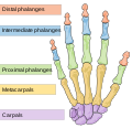
Intermetacarpal joints

Last updated
Intermetacarpal joints
Details
Identifiers
Latin articulationes intermetacarpales
TA98 A03.5.11.401
TA2 1831
FMA 71363
Anatomical terminology

The intermetacarpal joints are in the hand formed between the metacarpal bones. The bases of the second, third, fourth and fifth metacarpal bones articulate with one another by small surfaces covered with cartilage. The metacarpal bones are connected together by dorsal, palmar, and interosseous ligaments.

Contents

The synovial membrane for these joints is continuous with that of the carpometacarpal joints.

Additional images

See also

Related Research Articles

<span class="mw-page-title-main">Carpal bones</span> Eight small bones that make up the wrist (or carpus) that connects the hand to the forearm

The carpal bones are the eight small bones that make up the wrist (carpus) that connects the hand to the forearm. The term "carpus" and "carpal" is derived from the Latin carpus and the Greek καρπός (karpós), meaning "wrist". In human anatomy, the main role of the carpal bones is to articulate with the radial and ulnar heads to form a highly mobile condyloid joint, to provide attachments for thenar and hypothenar muscles, and to form part of the rigid carpal tunnel which allows the median nerve and tendons of the anterior forearm muscles to be transmitted to the hand and fingers.

<span class="mw-page-title-main">Wrist</span> Part of the arm between the lower arm and the hand

In human anatomy, the wrist is variously defined as (1) the carpus or carpal bones, the complex of eight bones forming the proximal skeletal segment of the hand; (2) the wrist joint or radiocarpal joint, the joint between the radius and the carpus and; (3) the anatomical region surrounding the carpus including the distal parts of the bones of the forearm and the proximal parts of the metacarpus or five metacarpal bones and the series of joints between these bones, thus referred to as wrist joints. This region also includes the carpal tunnel, the anatomical snuff box, bracelet lines, the flexor retinaculum, and the extensor retinaculum.

<span class="mw-page-title-main">Trapezoid bone</span> Carpal (wrist) bone

The trapezoid bone is a carpal bone in tetrapods, including humans. It is the smallest bone in the distal row of carpal bones that give structure to the palm of the hand. It may be known by its wedge-shaped form, the broad end of the wedge constituting the dorsal, the narrow end the palmar surface; and by its having four articular facets touching each other, and separated by sharp edges. It is homologous with the "second distal carpal" of reptiles and amphibians.

<span class="mw-page-title-main">Trapezium (bone)</span> Bone of the wrist

The trapezium bone is a carpal bone in the hand. It forms the radial border of the carpal tunnel.

<span class="mw-page-title-main">Metacarpal bones</span> Bones of hand

In human anatomy, the metacarpal bones or metacarpus, also known as the "palm bones", are the appendicular bones that form the intermediate part of the hand between the phalanges (fingers) and the carpal bones, which articulate with the forearm. The metacarpal bones are homologous to the metatarsal bones in the foot.

<span class="mw-page-title-main">Capitate bone</span> Carpal bone in the wrist

The capitate bone is a bone in the human wrist found in the center of the carpal bone region, located at the distal end of the radius and ulna bones. It articulates with the third metacarpal bone and forms the third carpometacarpal joint. The capitate bone is the largest of the carpal bones in the human hand. It presents, above, a rounded portion or head, which is received into the concavity formed by the scaphoid and lunate bones; a constricted portion or neck; and below this, the body. The bone is also found in many other mammals, and is homologous with the "third distal carpal" of reptiles and amphibians.

<span class="mw-page-title-main">Adductor pollicis muscle</span> Muscle in the thenar compartment

In human anatomy, the adductor pollicis muscle is a muscle in the hand that functions to adduct the thumb. It has two heads: transverse and oblique.

<span class="mw-page-title-main">Dorsal interossei of the foot</span> Four muscles situated between the metatarsal bones

In human anatomy, the dorsal interossei of the foot are four muscles situated between the metatarsal bones.

<span class="mw-page-title-main">Palmar interossei muscles</span> Muscles between the metacarpals

In human anatomy, the palmar or volar interossei are four muscles, one on the thumb that is occasionally missing, and three small, unipennate, central muscles in the hand that lie between the metacarpal bones and are attached to the index, ring, and little fingers. They are smaller than the dorsal interossei of the hand.

<span class="mw-page-title-main">Carpometacarpal joint</span>

The carpometacarpal (CMC) joints are five joints in the wrist that articulate the distal row of carpal bones and the proximal bases of the five metacarpal bones.

<span class="mw-page-title-main">Dorsal interossei of the hand</span> Muscles between the metacarpals

In human anatomy, the dorsal interossei (DI) are four muscles in the back of the hand that act to abduct (spread) the index, middle, and ring fingers away from hand's midline and assist in flexion at the metacarpophalangeal joints and extension at the interphalangeal joints of the index, middle and ring fingers.

<span class="mw-page-title-main">Palmar aponeurosis</span>

The palmar aponeurosis invests the muscles of the palm, and consists of central, lateral, and medial portions.

<span class="mw-page-title-main">First metacarpal bone</span>

The first metacarpal bone or the metacarpal bone of the thumb is the first bone proximal to the thumb. It is connected to the trapezium of the carpus at the first carpometacarpal joint and to the proximal thumb phalanx at the first metacarpophalangeal joint.

<span class="mw-page-title-main">Deep transverse metacarpal ligament</span> Ligament that connects the palmar surfaces of the heads of the second to fifth metacarpal bones

The deep transverse metacarpal ligament connects the palmar surfaces of metacarpophalangeal joints of all the fingers of the hand except the thumb.

<span class="mw-page-title-main">Intermetatarsal joints</span>

The intermetatarsal joints are the articulations between the base of metatarsal bones.

<span class="mw-page-title-main">Intercarpal joints</span> Joints of the carpal bones of the wrist

The intercarpal joints can be subdivided into three sets of joints : Those of the proximal row of carpal bones, those of the distal row of carpal bones, and those of the two rows with each other.

<span class="mw-page-title-main">Palmar carpometacarpal ligaments</span>

The Palmar carpometacarpal ligaments are a series of bands on the palmar surface of the carpometacarpal joints that connect the carpal bones to the second through fifth metacarpal bones. The second metacarpal is connected to the trapezium. The third metacarpal is connected to the trapezium, to the capitate, and to the hamate. The fourth and fifth metacarpals are connected to the hamate.

<span class="mw-page-title-main">Collateral ligaments of metacarpophalangeal joints</span>

In human anatomy, the radial (RCL) and ulnar (UCL) collateral ligaments of the metacarpophalangeal joints (MCP) of the hand are the primary stabilisers of the MCP joints. A collateral ligament flanks each MCP joint - one on either side. Each attaches proximally at the head of the metacarpal bone, and distally at the base of the phalynx. Each extends obliquely in a palmar direction from its proximal attachment to its distal attachment. The collateral ligaments allow spreading our the fingers with an open hand but not with the hand closed into a fist.

<span class="mw-page-title-main">Palmar plate</span>

In the human hand, palmar or volar plates are found in the metacarpophalangeal (MCP) and interphalangeal (IP) joints, where they reinforce the joint capsules, enhance joint stability, and limit hyperextension. The plates of the MCP and IP joints are structurally and functionally similar, except that in the MCP joints they are interconnected by a deep transverse ligament. In the MCP joints, they also indirectly provide stability to the longitudinal palmar arches of the hand. The volar plate of the thumb MCP joint has a transverse longitudinal rectangular shape, shorter than those in the fingers.

<span class="mw-page-title-main">Hand</span> Extremity at the end of an arm or forelimb

A hand is a prehensile, multi-fingered appendage located at the end of the forearm or forelimb of primates such as humans, chimpanzees, monkeys, and lemurs. A few other vertebrates such as the koala are often described as having "hands" instead of paws on their front limbs. The raccoon is usually described as having "hands" though opposable thumbs are lacking.

References

PD-icon.svgThis article incorporates text in the public domain from page 331 of the 20th edition of Gray's Anatomy (1918)