Interosseous membrane of forearm

Last updated
Interosseous membrane of forearm
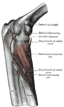
Gray420.png
Image of human elbow and forearm. Interosseous membrane labeled at bottom center.
Details
Identifiers
Latin membrana interossea antebrachii
TA98 A03.5.06.002
TA2 1761
FMA 23706
Anatomical terminology

The interosseous membrane of the forearm (rarely middle or intermediate radioulnar joint) is a fibrous sheet that connects the interosseous margins of the radius and the ulna. It is the main part of the radio-ulnar syndesmosis, a fibrous joint between the two bones.

Contents

Function

The interosseous membrane divides the forearm into anterior and posterior compartments, serves as a site of attachment for muscles of the forearm, and transfers loads placed on the forearm.

The interosseous membrane is designed to shift compressive loads (as in doing a hand-stand) from the distal radius to the proximal ulna. The fibers within the interosseous membrane are oriented obliquely so that when force is applied the fibers are drawn taut, shifting more of the load to the ulna. This reduces the wear and tear of placing the whole load on a single joint. [1] The role of the membrane in load shifting is illustrated when the interosseous membrane is cut; the forces on each bone equalize from their natural proportions. [2]

Additionally, as the forearm moves from pronation to supination, the interosseous membrane fibers change from a relaxed state, to a tense state in the neutral position. They once again become relaxed as the forearm enters pronation.

The interosseous membrane is composed of five ligaments:

  1. - Central band (key portion to be reconstructed in case of injury)
  2. - Accessory band
  3. - Distal oblique bundle
  4. - Proximal oblique cord
  5. - Dorsal oblique accessory cord

Injury

The interosseous membrane is composed of 5 ligaments:- Central band (key portion to be reconstructed in case of injury)- Accessory band- Distal oblique bundle- Proximal oblique cord- Dorsal oblique accessory cord 5 ligaments of interosseous membrane of forearm.png
The interosseous membrane is composed of 5 ligaments:- Central band (key portion to be reconstructed in case of injury)- Accessory band- Distal oblique bundle- Proximal oblique cord- Dorsal oblique accessory cord

Severe forearm injuries involving separation of the radius and ulna may be due to rupture of the interosseous membrane. Rupture can lead to proximal migration of the radius and an apparent lengthening of the ulna at the wrist. Often interosseous membrane tears are associated with adverse impacts on forearm rotation. MRI-assisted diagnosis has been used for mid-substance tears of the interosseous membrane but is expensive and not widely available. On the contrary, ultrasound has proven useful in diagnosing tears of the tibiofibular interosseous membrane of the leg, and this technique may also be applied to acute cases of membrane tears in the forearm due to its low cost and portability. [3]

See also

Related Research Articles

<span class="mw-page-title-main">Ulna</span> Medial bone from forearm

The ulna is a long bone found in the forearm that stretches from the elbow to the wrist, and when in anatomical position, is found on the medial side of the forearm. That is, the ulna is on the same side of the forearm as the little finger. It runs parallel to the radius, the other long bone in the forearm. The ulna is usually slightly longer than the radius, but the radius is thicker. Therefore, the radius is considered to be the larger of the two.

<span class="mw-page-title-main">Humerus</span> Long bone of the upper arm

The humerus is a long bone in the arm that runs from the shoulder to the elbow. It connects the scapula and the two bones of the lower arm, the radius and ulna, and consists of three sections. The humeral upper extremity consists of a rounded head, a narrow neck, and two short processes. The body is cylindrical in its upper portion, and more prismatic below. The lower extremity consists of 2 epicondyles, 2 processes, and 3 fossae. As well as its true anatomical neck, the constriction below the greater and lesser tubercles of the humerus is referred to as its surgical neck due to its tendency to fracture, thus often becoming the focus of surgeons.

<span class="mw-page-title-main">Brachioradialis</span> Muscle of the upper limb

The brachioradialis is a muscle of the forearm that flexes the forearm at the elbow. It is also capable of both pronation and supination, depending on the position of the forearm. It is attached to the distal styloid process of the radius by way of the brachioradialis tendon, and to the lateral supracondylar ridge of the humerus.

The forearm is the region of the upper limb between the elbow and the wrist. The term forearm is used in anatomy to distinguish it from the arm, a word which is used to describe the entire appendage of the upper limb, but which in anatomy, technically, means only the region of the upper arm, whereas the lower "arm" is called the forearm. It is homologous to the region of the leg that lies between the knee and the ankle joints, the crus.

<span class="mw-page-title-main">Radius (bone)</span> One of the two long bones of the forearm

The radius or radial bone is one of the two large bones of the forearm, the other being the ulna. It extends from the lateral side of the elbow to the thumb side of the wrist and runs parallel to the ulna. The ulna is usually slightly longer than the radius, but the radius is thicker. Therefore the radius is considered to be the larger of the two. It is a long bone, prism-shaped and slightly curved longitudinally.

<span class="mw-page-title-main">Upper limb</span> Consists of the arm, forearm, and hand

The upper limbs or upper extremities are the forelimbs of an upright-postured tetrapod vertebrate, extending from the scapulae and clavicles down to and including the digits, including all the musculatures and ligaments involved with the shoulder, elbow, wrist and knuckle joints. In humans, each upper limb is divided into the arm, forearm and hand, and is primarily used for climbing, lifting and manipulating objects.

In human anatomy, the extensor pollicis longus muscle (EPL) is a skeletal muscle located dorsally on the forearm. It is much larger than the extensor pollicis brevis, the origin of which it partly covers and acts to stretch the thumb together with this muscle.

The flexor pollicis longus is a muscle in the forearm and hand that flexes the thumb. It lies in the same plane as the flexor digitorum profundus. This muscle is unique to humans, being either rudimentary or absent in other primates. A meta-analysis indicated accessory flexor pollicis longus is present in around 48% of the population.

<span class="mw-page-title-main">Supinator muscle</span>

In human anatomy, the supinator is a broad muscle in the posterior compartment of the forearm, curved around the upper third of the radius. Its function is to supinate the forearm.

<span class="mw-page-title-main">Posterior interosseous nerve</span>

The posterior interosseous nerve is a nerve in the forearm. It is the continuation of the deep branch of the radial nerve, after this has crossed the supinator muscle. It is considerably diminished in size compared to the deep branch of the radial nerve. The nerve fibers originate from cervical segments C7 and C8 in the spinal column.

<span class="mw-page-title-main">Annular ligament of radius</span>

The annular ligament is a strong band of fibers that encircles the head of the radius, and retains it in contact with the radial notch of the ulna.

<span class="mw-page-title-main">Oblique cord</span>

The oblique cord is a ligament between the ulnar and radius bones in the forearm near the elbow. It takes the form of a small, flattened band, extending distally and laterally, from the lateral side of the ulnar tuberosity at the base of the coronoid process to the radius a little below the radial tuberosity. Its fibers run in the opposite direction to those of the Interosseous membrane of the forearm.

<span class="mw-page-title-main">Proximal radioulnar articulation</span>

The proximal radioulnar articulation, also known as the proximal radioulnar joint (PRUJ), is a synovial pivot joint between the circumference of the head of the radius and the ring formed by the radial notch of the ulna and the annular ligament.

<span class="mw-page-title-main">Anterior interosseous nerve</span>

The anterior interosseous nerve is a branch of the median nerve that supplies the deep muscles on the anterior of the forearm, except the ulnar (medial) half of the flexor digitorum profundus. Its nerve roots come from C8 and T1.

<span class="mw-page-title-main">Triangular fibrocartilage</span> Anatomical feature in the wrist

The triangular fibrocartilage complex (TFCC) is formed by the triangular fibrocartilage discus (TFC), the radioulnar ligaments (RULs) and the ulnocarpal ligaments (UCLs).

The posterior compartment of the forearm contains twelve muscles which primarily extend the wrist and digits. It is separated from the anterior compartment by the interosseous membrane between the radius and ulna.

<span class="mw-page-title-main">Elbow</span> Joint between the upper and lower parts of the arm

The elbow is the region between the upper arm and the forearm that surrounds the elbow joint. The elbow includes prominent landmarks such as the olecranon, the cubital fossa, and the lateral and the medial epicondyles of the humerus. The elbow joint is a hinge joint between the arm and the forearm; more specifically between the humerus in the upper arm and the radius and ulna in the forearm which allows the forearm and hand to be moved towards and away from the body. The term elbow is specifically used for humans and other primates, and in other vertebrates forelimb plus joint is used.

The Essex-Lopresti fracture is a fracture of the radial head of the forearm with concomitant dislocation of the distal radio-ulnar joint along with disruption of the thin interosseous membrane which holds them together. The injury is named after Peter Essex-Lopresti who described it in 1951.

<span class="mw-page-title-main">Extrinsic extensor muscles of the hand</span>

The extrinsic extensor muscles of the hand are located in the back of the forearm and have long tendons connecting them to bones in the hand, where they exert their action. Extrinsic denotes their location outside the hand. Extensor denotes their action which is to extend, or open flat, joints in the hand. They include the extensor carpi radialis longus (ECRL), extensor carpi radialis brevis (ECRB), extensor digitorum (ED), extensor digiti minimi (EDM), extensor carpi ulnaris (ECU), abductor pollicis longus (APL), extensor pollicis brevis (EPB), extensor pollicis longus (EPL), and extensor indicis (EI).

<span class="mw-page-title-main">Muscles of the thumb</span>

The muscles of the thumb are nine skeletal muscles located in the hand and forearm. The muscles allow for flexion, extension, adduction, abduction and opposition of the thumb. The muscles acting on the thumb can be divided into two groups: The extrinsic hand muscles, with their muscle bellies located in the forearm, and the intrinsic hand muscles, with their muscles bellies located in the hand proper.

References

  1. Saladin, Kenneth (2012). Anatomy and Physiology: The Unity of Form and Function 6th Edition. New York, NY: McGraw-Hill. p. 263. ISBN   978-0-07-337825-1.
  2. Birkbeck, D. P.; Failla, J. M.; Hoshaw, S. J.; Fyhrie, D. P.; Schaffler, M. (1997-11-01). "The interosseous membrane affects load distribution in the forearm". The Journal of Hand Surgery. 22 (6): 975–980. doi:10.1016/S0363-5023(97)80035-4. ISSN   0363-5023. PMID   9471063.
  3. Wallace, A. L.; Walsh, W. R.; van Rooijen, M.; Hughes, J. S.; Sonnabend, D. H. (1997-05-01). "The interosseous membrane in radioulnar dissociation". The Journal of Bone and Joint Surgery. British Volume. 79 (3): 422–427. doi: 10.1302/0301-620x.79b3.7142 . ISSN   0301-620X. PMID   9180321.