The carpal bones are the eight small bones that make up the wrist (carpus) that connects the hand to the forearm. The term "carpus" and "carpal" is derived from the Latin carpus and the Greek καρπός (karpós), meaning "wrist". In human anatomy, the main role of the carpal bones is to articulate with the radial and ulnar heads to form a highly mobile condyloid joint, to provide attachments for thenar and hypothenar muscles, and to form part of the rigid carpal tunnel which allows the median nerve and tendons of the anterior forearm muscles to be transmitted to the hand and fingers.

The ulna or ulnal bone is a long bone found in the forearm that stretches from the elbow to the wrist, and when in anatomical position, is found on the medial side of the forearm. That is, the ulna is on the same side of the forearm as the little finger. It runs parallel to the radius, the other long bone in the forearm. The ulna is longer and the radius is shorter, but the radius is thicker and the ulna is thinner. Therefore, the ulna is considered to be the smaller bone of the two bones in the lower arm. The corresponding bone in the lower leg is the fibula.

In human anatomy, the wrist is variously defined as (1) the carpus or carpal bones, the complex of eight bones forming the proximal skeletal segment of the hand; (2) the wrist joint or radiocarpal joint, the joint between the radius and the carpus and; (3) the anatomical region surrounding the carpus including the distal parts of the bones of the forearm and the proximal parts of the metacarpus or five metacarpal bones and the series of joints between these bones, thus referred to as wrist joints. This region also includes the carpal tunnel, the anatomical snuff box, bracelet lines, the flexor retinaculum, and the extensor retinaculum.

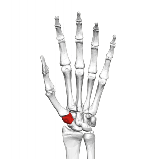
The trapezoid bone is a carpal bone in tetrapods, including humans. It is the smallest bone in the distal row of carpal bones that give structure to the palm of the hand. It may be known by its wedge-shaped form, the broad end of the wedge constituting the dorsal, the narrow end the palmar surface; and by its having four articular facets touching each other, and separated by sharp edges. It is homologous with the "second distal carpal" of reptiles and amphibians.
The trapezium bone is a carpal bone in the hand. It forms the radial border of the carpal tunnel.

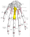
In human anatomy, the metacarpal bones or metacarpus, also known as the "palm bones", are the appendicular bones that form the intermediate part of the hand between the phalanges (fingers) and the carpal bones, which articulate with the forearm. The metacarpal bones are homologous to the metatarsal bones in the foot.

The scaphoid bone is one of the carpal bones of the wrist. It is situated between the hand and forearm on the thumb side of the wrist. It forms the radial border of the carpal tunnel. The scaphoid bone is the largest bone of the proximal row of wrist bones, its long axis being from above downward, lateralward, and forward. It is approximately the size and shape of a medium cashew nut.

The capitate bone is a bone in the human wrist found in the center of the carpal bone region, located at the distal end of the radius and ulna bones. It articulates with the third metacarpal bone and forms the third carpometacarpal joint. The capitate bone is the largest of the carpal bones in the human hand. It presents, above, a rounded portion or head, which is received into the concavity formed by the scaphoid and lunate bones; a constricted portion or neck; and below this, the body. The bone is also found in many other mammals, and is homologous with the "third distal carpal" of reptiles and amphibians.

The hamate bone, or unciform bone, Latin os hamatum and occasionally abbreviated as just hamatum, is a bone in the human wrist readily distinguishable by its wedge shape and a hook-like process ("hamulus") projecting from its palmar surface.

The lunate bone is a carpal bone in the human hand. It is distinguished by its deep concavity and crescentic outline. It is situated in the center of the proximal row carpal bones, which lie between the ulna and radius and the hand. The lunate carpal bone is situated between the lateral scaphoid bone and medial triquetral bone.

The radius or radial bone is one of the two large bones of the forearm, the other being the ulna. It extends from the lateral side of the elbow to the thumb side of the wrist and runs parallel to the ulna. The ulna is longer than the radius, but the radius is thicker. The radius is a long bone, prism-shaped and slightly curved longitudinally.

The triquetral bone is located in the wrist on the medial side of the proximal row of the carpus between the lunate and pisiform bones. It is on the ulnar side of the hand, but does not directly articulate with the ulna. Instead, it is connected to and articulates with the ulna through the Triangular fibrocartilage disc and ligament, which forms part of the ulnocarpal joint capsule. It connects with the pisiform, hamate, and lunate bones. It is the 2nd most commonly fractured carpal bone.
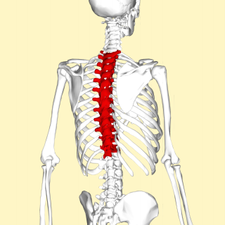
In vertebrates, thoracic vertebrae compose the middle segment of the vertebral column, between the cervical vertebrae and the lumbar vertebrae. In humans, there are twelve thoracic vertebrae and they are intermediate in size between the cervical and lumbar vertebrae; they increase in size going towards the lumbar vertebrae, with the lower ones being much larger than the upper. They are distinguished by the presence of facets on the sides of the bodies for articulation with the heads of the ribs, as well as facets on the transverse processes of all, except the eleventh and twelfth, for articulation with the tubercles of the ribs. By convention, the human thoracic vertebrae are numbered T1–T12, with the first one (T1) located closest to the skull and the others going down the spine toward the lumbar region.

The carpometacarpal (CMC) joints are five joints in the wrist that articulate the distal row of carpal bones and the proximal bases of the five metacarpal bones.

The fifth metacarpal bone is the most medial and second-shortest of the metacarpal bones.
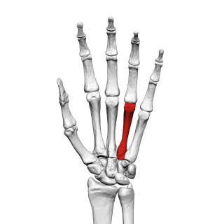
The fourth metacarpal bone is shorter and smaller than the third.
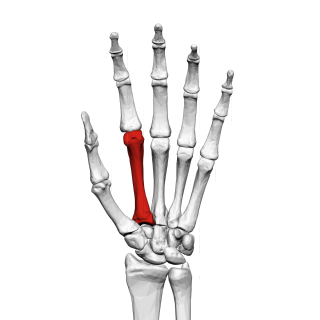
The second metacarpal bone is the longest, and its base the largest, of all the metacarpal bones.
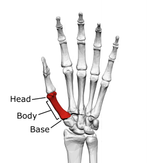
The first metacarpal bone or the metacarpal bone of the thumb is the first bone proximal to the thumb. It is connected to the trapezium of the carpus at the first carpometacarpal joint and to the proximal thumb phalanx at the first metacarpophalangeal joint.

The fourth metatarsal bone is a long bone in the foot. It is smaller in size than the third metatarsal bone and is the third longest of the five metatarsal bones. The fourth metatarsal is analogous to the fourth metacarpal bone in the hand
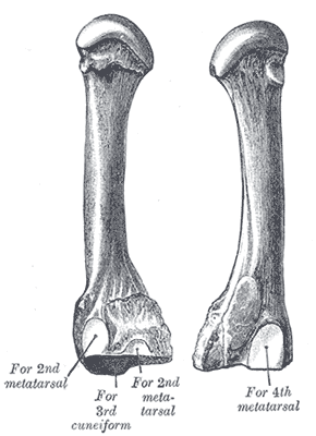
The third metatarsal bone is a long bone in the foot. It is the second longest metatarsal. The longest being the second metatarsal. The third metatarsal is analogous to the third metacarpal bone in the hand