Suprascapular notch

Last updated
Suprascapular notch
Suprascapular notch of left scapula03.png
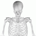
Costal surface of left scapula. Suprascapular notch shown in red.
Scapula ant - Suprascapular notch.png
Costal surface of left scapula. Suprascapular notch visible in the red square.
Details
Identifiers
Latin incisura scapulae
TA98 A02.4.01.015
TA2 1158
FMA 23236
Anatomical terms of bone

The suprascapular notch (or scapular notch) is a notch in the superior border of the scapula, just medial to the base of the coracoid process. [1] It is converted into the suprascapular canal by the suprascapular ligament. [2]

Contents

Structure

This notch is converted into a foramen by the suprascapular ligament, and serves for the passage of the suprascapular nerve. [2] The suprascapular vessels vary in number as well as in their course as they run at the suprascapular notch site. The suprascapular artery pass above the suprascapular ligament in most cases. The suprascapular vein may pass through the suprascapular notch or it may instead pass superior to the suprascapular ligament. [3] [1]

Types

Two main classification systems exist; others are modified approaches of the same principle.

Typing based on subjective observation of the suprascapular notch shape.

Introduced by Hrdicka 1942 and modified by Rengachary et al. 1979 There are six basic types of scapular notch:

Typing based on parametric measurements of depth to upper width ratio of the suprascapular notch

Introduced by Natsis et al. 2007 and modified by Polguj et al. 2011 There are five basic types of scapular notch:

The second method of suprascapular notch typing yields more practical approach in clinical diagnosis of the suprascapular nerve entrapment. [5]

Clinical significance

As the suprascapular nerve passes through the suprascapular notch, it is a common site of entrapment for the nerve. [1] [6] [7]

Suprascapular notch stenosis is a narrowing of the notch internal space that can potentially compress the suprascapular nerve leading to suprascapular nerve entrapment. Al-Redouan et al. 2020 predicted the morphological pattern of the suprascapular notch stenosis revealing higher incidence in the discrete notch (Type V according to the parametric measurements typing system). There are two main suprascapular stenosis patterns: [5]

The suprascapular nerve predictably passes through the suprascapular notch, so it is a good place for a local nerve block of the entire nerve. [8] [9]

Additional images

See also

Related Research Articles

<span class="mw-page-title-main">Scapula</span> Bone that connects the humerus (upper arm bone) with the clavicle (collar bone)

The scapula, also known as the shoulder blade, is the bone that connects the humerus with the clavicle. Like their connected bones, the scapulae are paired, with each scapula on either side of the body being roughly a mirror image of the other. The name derives from the Classical Latin word for trowel or small shovel, which it was thought to resemble.

<span class="mw-page-title-main">Coracoid process</span> Small hook-like structure on the lateral edge of the superior anterior portion of the scapula

The coracoid process is a small hook-like structure on the lateral edge of the superior anterior portion of the scapula. Pointing laterally forward, it, together with the acromion, serves to stabilize the shoulder joint. It is palpable in the deltopectoral groove between the deltoid and pectoralis major muscles.

<span class="mw-page-title-main">Ulnar nerve</span> Nerve which runs near the ulna bone

In human anatomy, the ulnar nerve is a nerve that runs near the ulna bone. The ulnar collateral ligament of elbow joint is in relation with the ulnar nerve. The nerve is the largest in the human body unprotected by muscle or bone, so injury is common. This nerve is directly connected to the little finger, and the adjacent half of the ring finger, innervating the palmar aspect of these fingers, including both front and back of the tips, perhaps as far back as the fingernail beds.

<span class="mw-page-title-main">Dorsal scapular nerve</span> Branch of the brachial plexus that supplies rhomboid muscles and levator scapulae

The dorsal scapular nerve is a branch of the brachial plexus, usually derived from the ventral ramus of cervical nerve C5. It provides motor innervation to the rhomboid major muscle, rhomboid minor muscle, and levator scapulae muscle.

Pudendal nerve entrapment (PNE), also known as Alcock canal syndrome, is an uncommon source of chronic pain in which the pudendal nerve is entrapped or compressed in Alcock's canal. There are several different types of PNE based on the site of entrapment anatomically. Pain is positional and is worsened by sitting. Other symptoms include genital numbness, fecal incontinence and urinary incontinence.

<span class="mw-page-title-main">Suprascapular nerve</span> Mixed nerve of the upper limb

The suprascapular nerve is a mixed nerve that branches from the upper trunk of the brachial plexus. It is derived from the ventral rami of cervical nerves C5-C6. It provides motor innervation to the supraspinatus muscle, and the infraspinatus muscle.

<span class="mw-page-title-main">Obturator internus muscle</span> One of six small hip muscles in the lateral rotator group

The internal obturator muscle or obturator internus muscle originates on the medial surface of the obturator membrane, the ischium near the membrane, and the rim of the pubis.

The posterior tibial artery of the lower limb is an artery that carries blood to the posterior compartment of the leg and plantar surface of the foot. It branches from the popliteal artery via the tibial-fibular trunk.

<span class="mw-page-title-main">Teres major muscle</span> Muscle of the upper limb

The teres major muscle is a muscle of the upper limb. It attaches to the scapula and the humerus and is one of the seven scapulohumeral muscles. It is a thick but somewhat flattened muscle.

<span class="mw-page-title-main">Transverse cervical artery</span> Artery of the neck

The transverse cervical artery is an artery in the neck and a branch of the thyrocervical trunk, running at a higher level than the suprascapular artery.

<span class="mw-page-title-main">Suprascapular artery</span> Artery of the neck

The suprascapular artery is a branch of the thyrocervical trunk on the neck.

<span class="mw-page-title-main">Mastoid part of the temporal bone</span> Back part of the sides of the skull base

The mastoid part of the temporal bone is the posterior (back) part of the temporal bone, one of the bones of the skull. Its rough surface gives attachment to various muscles and it has openings for blood vessels. From its borders, the mastoid part articulates with two other bones.

<span class="mw-page-title-main">Sternoclavicular joint</span> Joint between the manubrium of the sternum and the clavicle bone.

The sternoclavicular joint or sternoclavicular articulation is a synovial saddle joint between the manubrium of the sternum, and the clavicle, and the first costal cartilage. The joint possesses a joint capsule, and an articular disc, and is reinforced by multiple ligaments.

<span class="mw-page-title-main">Superior transverse scapular ligament</span>

The superior transverse ligament converts the suprascapular notch into a foramen or opening.

<span class="mw-page-title-main">Inferior transverse ligament of scapula</span>

The inferior transverse ligament is a weak membranous band, situated behind the neck of the scapula and stretching from the lateral border of the spine to the margin of the glenoid cavity.

<span class="mw-page-title-main">Spine of scapula</span> Bony plate on the scapula

The spine of the scapula or scapular spine is a prominent plate of bone, which crosses obliquely the medial four-fifths of the scapula at its upper part, and separates the supra- from the infraspinatous fossa.

The saphenous nerve is the largest cutaneous branch of the femoral nerve. It is derived from the lumbar plexus (L3-L4). It is a strictly sensory nerve, and has no motor function. It commences in the proximal (upper) thigh and travels along the adductor canal. Upon exiting the adductor canal, the saphenous nerve terminates by splitting into two terminal branches: the sartorial nerve, and the infrapatellar nerve. The saphenous nerve is responsible for providing sensory innervation to the skin of the anteromedial leg.

<span class="mw-page-title-main">Quadrangular space</span>

The quadrangular space, also known as the quadrilateral space [of Velpeau] and the foramen humerotricipitale, is one of the three spaces in the axillary space. The other two spaces are: triangular space and triangular interval.

<span class="mw-page-title-main">Suprascapular vein</span> Vein of the neck

The suprascapular vein is a vein running above the scapula. It drains into the external jugular vein. It drains the posterior region around the scapula.

<span class="mw-page-title-main">Suprascapular canal</span> Anatomical passage between two openings found on the upper dorsal aspect of the shoulder

The suprascapular canal is an anatomical passage between two openings found on the upper dorsal aspect of the shoulder. It is found bilaterally running on superior-lateral aspect of the dorsal surface of the scapula underneath the supraspinatus muscle.

References

  1. 1 2 3 Al-Redouan, Azzat; Holding, Keiv; Kachlik, David (2021). ""Suprascapular canal": Anatomical and topographical description and its clinical implication in entrapment syndrome". Annals of Anatomy. 233: 151593. doi: 10.1016/j.aanat.2020.151593 . PMID   32898658.
  2. 1 2 Nathan, Jay K.; McGillicuddy, John E. (2015-01-01), Tubbs, R. Shane; Rizk, Elias; Shoja, Mohammadali M.; Loukas, Marios (eds.), "Chapter 38 - Anatomy of the Ventral Rami, Upper Trunk, and Its Divisions and Branches", Nerves and Nerve Injuries, San Diego: Academic Press, pp. 527–535, doi:10.1016/b978-0-12-410390-0.00040-8, ISBN   978-0-12-410390-0 , retrieved 2020-10-19
  3. Polguj, Michał; Rożniecki, Jacek; Sibiński, Marcin; Grzegorzewski, Andrzej; Majos, Agata; Topol, Mirosław (2015). "The variable morphology of suprascapular nerve and vessels at suprascapular notch: a proposal for classification and its potential clinical implications". Knee Surg Sports Traumatol Arthrosc. 23 (5): 1542–1548. doi: 10.1007/s00167-014-2937-1 . PMC   4555201 . PMID   24633009.
  4. White, Tim D.; Black, Michael T.; Folkens, Pieter A. (2012-01-01), White, Tim D.; Black, Michael T.; Folkens, Pieter A. (eds.), "Chapter 8 - Shoulder Girdle: Clavicle and Scapula", Human Osteology (Third Edition), San Diego: Academic Press, pp. 161–174, doi:10.1016/b978-0-12-374134-9.50008-8, ISBN   978-0-12-374134-9 , retrieved 2020-10-19
  5. 1 2 Al-Redouan, Azzat; Hudak, Radovan; Nanka, Ondrej; Kachlik, David (2020). "The morphological stenosis pattern of the suprascapular notch is revealed yielding higher incidence in the discrete type and elucidating the inevitability of osteoplasty in horizontally oriented stenosis". Knee Surg Sports Traumatol Arthrosc. 29 (7): 2272–2280. doi:10.1007/s00167-020-06168-1. PMID   32712687. S2CID   220797681.
  6. Preston, David C.; Shapiro, Barbara E. (2013-01-01), Preston, David C.; Shapiro, Barbara E. (eds.), "31 - Proximal Neuropathies of the Shoulder and Arm", Electromyography and Neuromuscular Disorders (Third Edition), London: W.B. Saunders, pp. 487–500, doi:10.1016/b978-1-4557-2672-1.00031-3, ISBN   978-1-4557-2672-1 , retrieved 2020-10-19
  7. Bouche, P. (2013-01-01), Said, Gérard; Krarup, Christian (eds.), "Chapter 19 - Compression and entrapment neuropathies", Handbook of Clinical Neurology, Peripheral Nerve Disorders, Elsevier, 115: 311–366, doi:10.1016/b978-0-444-52902-2.00019-9, ISBN   9780444529022, PMID   23931789 , retrieved 2020-10-19
  8. Wilkinson, Laura J. "Defunct DOI". Crossref. doi: 10.1016/b978-032304184-3.50050-9 . Retrieved 2020-10-19.
  9. Molloy, Robert E. (2005-01-01), Benzon, Honorio T.; Raja, Srinivasa N.; Molloy, Robert E.; Liu, Spencer S. (eds.), "Chapter 75 - Truncal Blocks: Intercostal, Paravertebral, Interpleural, Suprascapular, Ilioinguinal, and Iliohypogastric Nerve Blocks", Essentials of Pain Medicine and Regional Anesthesia (Second Edition), Philadelphia: Churchill Livingstone, pp. 636–644, doi:10.1016/b978-0-443-06651-1.50079-4, ISBN   978-0-443-06651-1 , retrieved 2020-10-19

PD-icon.svgThis article incorporates text in the public domain from page 204 of the 20th edition of Gray's Anatomy (1918)