Radial tuberosity

Last updated
Radial tuberosity
Tuberositasradii.png
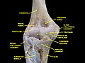
Left elbow-joint, showing anterior and ulnar collateral ligaments. (Radial tuberosity visible at center right.)
Gray213.png
Bones of left forearm. Anterior aspect. (Radius is bone on right. Radial tuberosity is visible at upper left of radius.)
Details
Identifiers
Latin tuberositas radii
TA98 A02.4.05.007
TA2 1216
FMA 23489
Anatomical terms of bone

Beneath the neck of the radius, on the medial side, is an eminence, the radial tuberosity; its surface is divided into:

Contents

Ligaments that support the elbow joint also attach to the radial tuberosity. [5]

Related Research Articles

<span class="mw-page-title-main">Humerus</span> Long bone of the upper arm

The humerus is a long bone in the arm that runs from the shoulder to the elbow. It connects the scapula and the two bones of the lower arm, the radius and ulna, and consists of three sections. The humeral upper extremity consists of a rounded head, a narrow neck, and two short processes. The body is cylindrical in its upper portion, and more prismatic below. The lower extremity consists of 2 epicondyles, 2 processes, and 3 fossae. As well as its true anatomical neck, the constriction below the greater and lesser tubercles of the humerus is referred to as its surgical neck due to its tendency to fracture, thus often becoming the focus of surgeons.

<span class="mw-page-title-main">Biceps</span> Muscle on the front of the upper arm

The biceps or biceps brachii are a large muscle that lies on the front of the upper arm between the shoulder and the elbow. Both heads of the muscle arise on the scapula and join to form a single muscle belly which is attached to the upper forearm. While the biceps crosses both the shoulder and elbow joints, its main function is at the elbow where it flexes the forearm and supinates the forearm. Both these movements are used when opening a bottle with a corkscrew: first biceps screws in the cork (supination), then it pulls the cork out (flexion).

<span class="mw-page-title-main">Brachioradialis</span> Muscle of the upper limb

The brachioradialis is a muscle of the forearm that flexes the forearm at the elbow. It is also capable of both pronation and supination, depending on the position of the forearm. It is attached to the distal styloid process of the radius by way of the brachioradialis tendon, and to the lateral supracondylar ridge of the humerus.

<span class="mw-page-title-main">Radial nerve</span> Nerve in the human body that supplies the posterior portion of the upper limb

The radial nerve is a nerve in the human body that supplies the posterior portion of the upper limb. It innervates the medial and lateral heads of the triceps brachii muscle of the arm, as well as all 12 muscles in the posterior osteofascial compartment of the forearm and the associated joints and overlying skin.

<span class="mw-page-title-main">Brachialis muscle</span> Flexor muscle in the upper arm

The brachialis is a muscle in the upper arm that flexes the elbow. It lies beneath the biceps brachii, and makes up part of the floor of the region known as the cubital fossa. It originates from the anterior aspect of the distal humerus; it inserts onto the tuberosity of the ulna. It is innervated by the musculocutaneous nerve, and commonly also receives additional innervation from the radial nerve. The brachialis is the prime mover of elbow flexion generating about 50% more power than the biceps.

<span class="mw-page-title-main">Ulnar nerve</span> Nerve which runs near the ulna bone

In human anatomy, the ulnar nerve is a nerve that runs near the ulna bone. The ulnar collateral ligament of elbow joint is in relation with the ulnar nerve. The nerve is the largest in the human body unprotected by muscle or bone, so injury is common. This nerve is directly connected to the little finger, and the adjacent half of the ring finger, innervating the palmar aspect of these fingers, including both front and back of the tips, perhaps as far back as the fingernail beds.

<span class="mw-page-title-main">Radius (bone)</span> One of the two long bones of the forearm

The radius or radial bone is one of the two large bones of the forearm, the other being the ulna. It extends from the lateral side of the elbow to the thumb side of the wrist and runs parallel to the ulna. The ulna is longer than the radius, but the radius is thicker. The radius is a long bone, prism-shaped and slightly curved longitudinally.

<span class="mw-page-title-main">Omohyoid muscle</span> Human neck muscle

The omohyoid muscle is a muscle in the neck. It is one of the infrahyoid muscles. It consists of two bellies separated by an intermediate tendon. Its inferior belly is attached to the scapula; its superior belly is attached to the hyoid bone. Its intermediate tendon is anchored to the clavicle and first rib by a fascial sling. The omohyoid is innervated by the ansa cervicalis of the cervical plexus. It acts to depress the hyoid bone.

<span class="mw-page-title-main">Triceps</span> Muscle on the back of the upper arm

The triceps, or triceps brachii, is a large muscle on the back of the upper limb of many vertebrates. It consists of 3 parts: the medial, lateral, and long head. It is the muscle principally responsible for extension of the elbow joint.

The flexor pollicis longus is a muscle in the forearm and hand that flexes the thumb. It lies in the same plane as the flexor digitorum profundus. This muscle is unique to humans, being either rudimentary or absent in other primates. A meta-analysis indicated accessory flexor pollicis longus is present in around 48% of the population.

<span class="mw-page-title-main">Supinator muscle</span> Muscle of the forearm in humans

In human anatomy, the supinator is a broad muscle in the posterior compartment of the forearm, curved around the upper third of the radius. Its function is to supinate the forearm.

<span class="mw-page-title-main">Fibularis tertius</span> Muscle of the human body located in the lower limb

In human anatomy, the fibularis tertius is a muscle in the anterior compartment of the leg. It acts to tilt the sole of the foot away from the midline of the body (eversion) and to pull the foot upward toward the body (dorsiflexion).

<span class="mw-page-title-main">Olecranon fossa</span> Deep triangular depression on the posterior end of the humerus

The olecranon fossa is a deep triangular depression on the posterior side of the humerus, superior to the trochlea. It provides space for the olecranon of the ulna during extension of the forearm.

<span class="mw-page-title-main">Radial collateral ligament of elbow joint</span> Ligament of elbow

The radial collateral ligament (RCL), lateral collateral ligament (LCL), or external lateral ligament is a ligament in the elbow on the side of the radius.

<span class="mw-page-title-main">Glenoid labrum</span> Structure on the shoulder blade

The glenoid labrum is a fibrocartilaginous structure attached around the rim of the glenoid cavity on the shoulder blade. The shoulder joint is considered a ball-and-socket joint. However, in bony terms the 'socket' is quite shallow and small, covering at most only a third of the 'ball'. The socket is deepened by the glenoid labrum, stabilizing the shoulder joint.

<span class="mw-page-title-main">Supraglenoid tubercle</span> Region of the scapula from which the long head of the biceps brachii muscle originates

The supraglenoid tubercle is a region of the scapula from which the long head of the biceps brachii muscle originates. It is a small, rough projection superior to the glenoid cavity near the base of the coracoid process. The term supraglenoid is from the Latin supra, meaning above, and glenoid, meaning socket or cavity.

<span class="mw-page-title-main">Radial styloid process</span> Bone projection on the radius

The radial styloid process is a projection of bone on the lateral surface of the distal radius bone.

<span class="mw-page-title-main">Tuberosity of the ulna</span>

The tuberosity of the ulna is a rough eminence on the proximal end of the ulna. It occurs at the junction of the antero-inferior surface of the coronoid process with the front of the body. It provides an insertion point to a tendon of the brachialis.

<span class="mw-page-title-main">Radial fossa</span> Groove on the distal end of the humerus

The radial fossa is a slight depression found on the humerus above the front part of the capitulum. It receives the anterior border of the head of the radius when the forearm is flexed.

<span class="mw-page-title-main">Head of radius</span> Bone of the arm

The head of the radius has a cylindrical form, and on its upper surface is a shallow cup or fovea for articulation with the capitulum of the humerus. The circumference of the head is smooth; it is broad medially where it articulates with the radial notch of the ulna, narrow in the rest of its extent, which is embraced by the annular ligament.

References

PD-icon.svgThis article incorporates text in the public domain from page 219 of the 20th edition of Gray's Anatomy (1918)

  1. "Radius". Anatomedia. Retrieved 18 October 2020.
  2. Metzler, A. V.; Greiwe, R. M. (2016-01-01), Greiwe, R. Michael (ed.), "3 - Management of acute and chronic distal biceps ruptures", Shoulder and Elbow Trauma and its Complications, Woodhead Publishing Series in Biomaterials, Woodhead Publishing, pp. 47–64, doi:10.1016/b978-1-78242-450-5.00003-4, ISBN   978-1-78242-450-5 , retrieved 2020-10-18
  3. Wachtel, Tom J., ed. (2007-01-01), "- T", Geriatric Clinical Advisor, Philadelphia: Mosby, pp. 204–221, doi:10.1016/b978-032304195-9.50019-5, ISBN   978-0-323-04195-9 , retrieved 2020-10-22
  4. Watkins, Jeffrey P. (2012-01-01), Auer, Jörg A.; Stick, John A. (eds.), "Chapter 95 - Radius and Ulna", Equine Surgery (Fourth Edition), Saint Louis: W.B. Saunders, pp. 1363–1378, doi:10.1016/b978-1-4377-0867-7.00095-8, ISBN   978-1-4377-0867-7 , retrieved 2020-10-22
  5. Dyson, Sue J. (2011-01-01), Ross, Mike W.; Dyson, Sue J. (eds.), "Chapter 40 - The Elbow, Brachium, and Shoulder", Diagnosis and Management of Lameness in the Horse (Second Edition), Saint Louis: W.B. Saunders, pp. 456–474, doi:10.1016/b978-1-4160-6069-7.00040-7, ISBN   978-1-4160-6069-7 , retrieved 2020-10-22

Additional images