
The humerus is a long bone in the arm that runs from the shoulder to the elbow. It connects the scapula and the two bones of the lower arm, the radius and ulna, and consists of three sections. The humeral upper extremity consists of a rounded head, a narrow neck, and two short processes. The body is cylindrical in its upper portion, and more prismatic below. The lower extremity consists of 2 epicondyles, 2 processes, and 3 fossae. As well as its true anatomical neck, the constriction below the greater and lesser tubercles of the humerus is referred to as its surgical neck due to its tendency to fracture, thus often becoming the focus of surgeons.

The brachioradialis is a muscle of the forearm that flexes the forearm at the elbow. It is also capable of both pronation and supination, depending on the position of the forearm. It is attached to the distal styloid process of the radius by way of the brachioradialis tendon, and to the lateral supracondylar ridge of the humerus.

The human shoulder is made up of three bones: the clavicle (collarbone), the scapula, and the humerus as well as associated muscles, ligaments and tendons. The articulations between the bones of the shoulder make up the shoulder joints. The shoulder joint, also known as the glenohumeral joint, is the major joint of the shoulder, but can more broadly include the acromioclavicular joint. In human anatomy, the shoulder joint comprises the part of the body where the humerus attaches to the scapula, and the head sits in the glenoid cavity. The shoulder is the group of structures in the region of the joint.

The nasal bones are two small oblong bones, varying in size and form in different individuals; they are placed side by side at the middle and upper part of the face and by their junction, form the bridge of the upper one third of the nose.
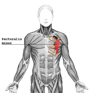
The pectoralis minor is a thin, triangular muscle, situated at the upper part of the chest, beneath the pectoralis major in the human body.

The internal pudendal artery is one of the three pudendal arteries that branches off the internal iliac artery, providing blood to the external genitalia.

The teres minor is a narrow, elongated muscle of the rotator cuff. The muscle originates from the lateral border and adjacent posterior surface of the corresponding right or left scapula and inserts at both the greater tubercle of the humerus and the posterior surface of the joint capsule.

The pectineus muscle is a flat, quadrangular muscle, situated at the anterior (front) part of the upper and medial (inner) aspect of the thigh. The pectineus muscle is the most anterior adductor of the hip. The muscle does adduct and internally rotate the thigh but its primary function is hip flexion.

The supraspinatus is a relatively small muscle of the upper back that runs from the supraspinous fossa superior portion of the scapula to the greater tubercle of the humerus. It is one of the four rotator cuff muscles and also abducts the arm at the shoulder. The spine of the scapula separates the supraspinatus muscle from the infraspinatus muscle, which originates below the spine.

In human anatomy, the infraspinatus muscle is a thick triangular muscle, which occupies the chief part of the infraspinatous fossa. As one of the four muscles of the rotator cuff, the main function of the infraspinatus is to externally rotate the humerus and stabilize the shoulder joint.

The shoulder joint is structurally classified as a synovial ball and socket joint and functionally as a diarthrosis and multiaxial joint. It involves articulation between the glenoid cavity of the scapula and the head of the humerus.

The rectus capitis posterior minor arises by a narrow pointed tendon from the tubercle on the posterior arch of the atlas, and, widening as it ascends, is inserted into the medial part of the inferior nuchal line of the occipital bone and the surface between it and the foramen magnum, and also takes some attachment to the spinal dura mater.

The teres major muscle is a muscle of the upper limb. It attaches to the scapula and the humerus and is one of the seven scapulohumeral muscles. It is a thick but somewhat flattened muscle.
The semimembranosus is the most medial of the three hamstring muscles. It is so named because it has a flat tendon of origin. It lies posteromedially in the thigh, deep to the semitendinosus.

The sacrotuberous ligament is situated at the lower and back part of the pelvis. It is flat, and triangular in form; narrower in the middle than at the ends.

The glenoid cavity or glenoid fossa of scapula is a part of the shoulder. It is a shallow, pyriform articular surface, which is located on the lateral angle of the scapula. It is directed laterally and forward and articulates with the head of the humerus; it is broader below than above and its vertical diameter is the longest.

The crest of the ilium is the superior border of the wing of ilium and the superiolateral margin of the greater pelvis.

The medial epicondyle of the humerus is an epicondyle of the humerus bone of the upper arm in humans. It is larger and more prominent than the lateral epicondyle and is directed slightly more posteriorly in the anatomical position. In birds, where the arm is somewhat rotated compared to other tetrapods, it is called the ventral epicondyle of the humerus. In comparative anatomy, the more neutral term entepicondyle is used.

The lesser tubercle of the humerus, although smaller, is more prominent than the greater tubercle: it is situated in front, and is directed medially and anteriorly.

In the vertebrate spinal column, each vertebra is an irregular bone with a complex structure composed of bone and some hyaline cartilage, the proportions of which vary according to the segment of the backbone and the species of vertebrate.


