Trapezium (bone)

Last updated
Trapezium
Trapezium bone (left)01 palmar view.png
Left hand anterior view (palmar view). Trapezium shown in red.
Gray225.png
The left trapezium bone ("navicular" refers to scaphoid, "lesser multangular" refers to trapezoid)
Details
Articulations 1st metacarpal distally
scaphoid proximally
trapezoid medially
2nd metacarpal medially
Identifiers
Latin os trapezium, os multangulum majus
MeSH D051222
TA98 A02.4.08.008
TA2 1255
FMA 23721
Anatomical terms of bone

The trapezium bone (greater multangular bone) is a carpal bone in the hand. It forms the radial border of the carpal tunnel.

Contents

Structure

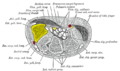
The trapezium is distinguished by a deep groove on its anterior surface. It is situated at the radial side of the carpus, between the scaphoid and the first metacarpal bone (the metacarpal bone of the thumb). It is homologous with the first distal carpal of reptiles and amphibians.

Surfaces

The trapezium is an irregular-shaped carpal bone found within the hand. The trapezium is found within the distal row of carpal bones, and is directly adjacent to the metacarpal bone of the thumb. On its ulnar surface are found the trapezoid and scaphoid bones. [1] :708

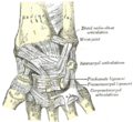
The superior surface is directed upward and medialward; medially it is smooth, and articulates with the scaphoid; laterally it is rough and continuous with the lateral surface.

The inferior surface is oval, concave from side to side, convex from before backward, so as to form a saddle-shaped surface for articulation with the base of the first metacarpal bone. This saddle-shaped articulation is partially responsible for the thumb's opposable motion.

The dorsal surface is smooth.

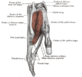
The palmar surface is narrow and rough. At its upper part is a deep groove, running from above obliquely downward and medialward; it transmits the tendon of the Flexor carpi radialis, and is bounded laterally by an oblique ridge. This surface gives origin to the Opponens pollicis and to the Abductor and Flexor pollicis brevis; it also affords attachment to the transverse carpal ligament.

The lateral surface is broad and rough, for the attachment of ligaments.

The medial surface presents two facets; the upper, large and concave, articulates with the trapezoid bone; the lower, small and oval, with the base of the second metacarpal.

Tubercle of trapezium

The tubercle of trapezium is a tubercle found on the anterior surface of the bone. [1] :708 It is where sometimes abductor pollicis brevis muscle attaches.

Function

The carpal bones function as a unit to provide a bony superstructure for the hand. The trapezium is the most radial of the bones surrounding the carpal tunnel. [1] :708 It is important in thumb movement. [2]

Clinical relevance

The trapezium is susceptible to arthritis at the joint with the metacarpal bone of the thumb, due to overuse. [2]

History

The etymology derives from the Greek trapezion which means "a little table", from trapeza meaning "table", itself from (te)tra- "four" and pod- "foot". The name was first documented in 1840. [3]

Additional images

See also

Related Research Articles

<span class="mw-page-title-main">Carpal bones</span> Eight small bones that make up the wrist (or carpus) that connects the hand to the forearm

The carpal bones are the eight small bones that make up the wrist (carpus) that connects the hand to the forearm. The term "carpus" and "carpal" is derived from the Latin carpus and the Greek καρπός (karpós), meaning "wrist". In human anatomy, the main role of the carpal bones is to articulate with the radial and ulnar heads to form a highly mobile condyloid joint, to provide attachments for thenar and hypothenar muscles, and to form part of the rigid carpal tunnel which allows the median nerve and tendons of the anterior forearm muscles to be transmitted to the hand and fingers.

<span class="mw-page-title-main">Wrist</span> Part of the arm between the lower arm and the hand

In human anatomy, the wrist is variously defined as (1) the carpus or carpal bones, the complex of eight bones forming the proximal skeletal segment of the hand; (2) the wrist joint or radiocarpal joint, the joint between the radius and the carpus and; (3) the anatomical region surrounding the carpus including the distal parts of the bones of the forearm and the proximal parts of the metacarpus or five metacarpal bones and the series of joints between these bones, thus referred to as wrist joints. This region also includes the carpal tunnel, the anatomical snuff box, bracelet lines, the flexor retinaculum, and the extensor retinaculum.

<span class="mw-page-title-main">Anatomical snuffbox</span> Indent on back of hand between tendons

The anatomical snuff box or snuffbox or foveola radialis is a triangular deepening on the radial, dorsal aspect of the hand—at the level of the carpal bones, specifically, the scaphoid and trapezium bones forming the floor. The name originates from the use of this surface for placing and then sniffing powdered tobacco, or "snuff." It is sometimes referred to by its French name tabatière.

<span class="mw-page-title-main">Trapezoid bone</span> Carpal (wrist) bone

The trapezoid bone is a carpal bone in tetrapods, including humans. It is the smallest bone in the distal row of carpal bones that give structure to the palm of the hand. It may be known by its wedge-shaped form, the broad end of the wedge constituting the dorsal, the narrow end the palmar surface; and by its having four articular facets touching each other, and separated by sharp edges. It is homologous with the "second distal carpal" of reptiles and amphibians.

<span class="mw-page-title-main">Metacarpal bones</span> Bones of hand

In human anatomy, the metacarpal bones or metacarpus, also known as the "palm bones", are the appendicular bones that form the intermediate part of the hand between the phalanges (fingers) and the carpal bones, which articulate with the forearm. The metacarpal bones are homologous to the metatarsal bones in the foot.

<span class="mw-page-title-main">Scaphoid bone</span> Bone of the carpus

The scaphoid bone is one of the carpal bones of the wrist. It is situated between the hand and forearm on the thumb side of the wrist. It forms the radial border of the carpal tunnel. The scaphoid bone is the largest bone of the proximal row of wrist bones, its long axis being from above downward, lateralward, and forward. It is approximately the size and shape of a medium cashew nut.

<span class="mw-page-title-main">Capitate bone</span> Carpal bone in the wrist

The capitate bone is a bone in the human wrist found in the center of the carpal bone region, located at the distal end of the radius and ulna bones. It articulates with the third metacarpal bone and forms the third carpometacarpal joint. The capitate bone is the largest of the carpal bones in the human hand. It presents, above, a rounded portion or head, which is received into the concavity formed by the scaphoid and lunate bones; a constricted portion or neck; and below this, the body. The bone is also found in many other mammals, and is homologous with the "third distal carpal" of reptiles and amphibians.

<span class="mw-page-title-main">Hamate bone</span> Carpal bone in the wrist

The hamate bone, or unciform bone, Latin os hamatum and occasionally abbreviated as just hamatum, is a bone in the human wrist readily distinguishable by its wedge shape and a hook-like process ("hamulus") projecting from its palmar surface.

<span class="mw-page-title-main">Lunate bone</span> Carpal bone in the human hand

The lunate bone is a carpal bone in the human hand. It is distinguished by its deep concavity and crescentic outline. It is situated in the center of the proximal row carpal bones, which lie between the ulna and radius and the hand. The lunate carpal bone is situated between the lateral scaphoid bone and medial triquetral bone.

<span class="mw-page-title-main">Radius (bone)</span> One of the two long bones of the forearm

The radius or radial bone is one of the two large bones of the forearm, the other being the ulna. It extends from the lateral side of the elbow to the thumb side of the wrist and runs parallel to the ulna. The ulna is longer than the radius, but the radius is thicker. The radius is a long bone, prism-shaped and slightly curved longitudinally.

<span class="mw-page-title-main">Triquetral bone</span> Bone in the wrist

The triquetral bone is located in the wrist on the medial side of the proximal row of the carpus between the lunate and pisiform bones. It is on the ulnar side of the hand, but does not directly articulate with the ulna. Instead, it is connected to and articulates with the ulna through the Triangular fibrocartilage disc and ligament, which forms part of the ulnocarpal joint capsule. It connects with the pisiform, hamate, and lunate bones. It is the 2nd most commonly fractured carpal bone.

<span class="mw-page-title-main">Upper limb</span> Consists of the arm, forearm, and hand

The upper limbs or upper extremities are the forelimbs of an upright-postured tetrapod vertebrate, extending from the scapulae and clavicles down to and including the digits, including all the musculatures and ligaments involved with the shoulder, elbow, wrist and knuckle joints. In humans, each upper limb is divided into the arm, forearm and hand, and is primarily used for climbing, lifting and manipulating objects.

<span class="mw-page-title-main">Flexor pollicis brevis muscle</span> Muscle in the thenar compartment

The flexor pollicis brevis is a muscle in the hand that flexes the thumb. It is one of three thenar muscles. It has both a superficial part and a deep part.

In human anatomy, the abductor pollicis longus (APL) is one of the extrinsic muscles of the hand. Its major function is to abduct the thumb at the wrist. Its tendon forms the anterior border of the anatomical snuffbox.

<span class="mw-page-title-main">Carpometacarpal joint</span>

The carpometacarpal (CMC) joints are five joints in the wrist that articulate the distal row of carpal bones and the proximal bases of the five metacarpal bones.

<span class="mw-page-title-main">First metacarpal bone</span>

The first metacarpal bone or the metacarpal bone of the thumb is the first bone proximal to the thumb. It is connected to the trapezium of the carpus at the first carpometacarpal joint and to the proximal thumb phalanx at the first metacarpophalangeal joint.

<span class="mw-page-title-main">Midcarpal joint</span>

The midcarpal joint is formed by the scaphoid, lunate, and triquetral bones in the proximal row, and the trapezium, trapezoid, capitate, and hamate bones in the distal row. The distal pole of the scaphoid articulates with two trapezial bones as a gliding type of joint. The proximal end of the scaphoid combines with the lunate and triquetrum to form a deep concavity that articulates with the convexity of the combined capitate and hamate in a form of diarthrodial, almost condyloid joint.

<span class="mw-page-title-main">Hand</span> Extremity at the end of an arm or forelimb

A hand is a prehensile, multi-fingered appendage located at the end of the forearm or forelimb of primates such as humans, chimpanzees, monkeys, and lemurs. A few other vertebrates such as the koala are often described as having "hands" instead of paws on their front limbs. The raccoon is usually described as having "hands" though opposable thumbs are lacking.

<span class="mw-page-title-main">Extrinsic extensor muscles of the hand</span>

The extrinsic extensor muscles of the hand are located in the back of the forearm and have long tendons connecting them to bones in the hand, where they exert their action. Extrinsic denotes their location outside the hand. Extensor denotes their action which is to extend, or open flat, joints in the hand. They include the extensor carpi radialis longus (ECRL), extensor carpi radialis brevis (ECRB), extensor digitorum (ED), extensor digiti minimi (EDM), extensor carpi ulnaris (ECU), abductor pollicis longus (APL), extensor pollicis brevis (EPB), extensor pollicis longus (EPL), and extensor indicis (EI).

<span class="mw-page-title-main">Muscles of the thumb</span>

The muscles of the thumb are nine skeletal muscles located in the hand and forearm. The muscles allow for flexion, extension, adduction, abduction and opposition of the thumb. The muscles acting on the thumb can be divided into two groups: The extrinsic hand muscles, with their muscle bellies located in the forearm, and the intrinsic hand muscles, with their muscles bellies located in the hand proper.

References

  1. 1 2 3 Drake, Richard L.; Vogl, Wayne; Tibbitts, Adam W.M. Mitchell; illustrations by Richard; Richardson, Paul (2005). Gray's anatomy for students. Philadelphia: Elsevier/Churchill Livingstone. ISBN   978-0-8089-2306-0.
  2. 1 2 Eathorne, SW (Mar 2005). "The wrist: clinical anatomy and physical examination--an update". Primary Care. 32 (1): 17–33. doi:10.1016/j.pop.2004.11.009. PMID   15831311.
  3. Harper, Douglas. "Trapezium". Online Etymology Dictionary. Retrieved 5 January 2014.