The scapula, also known as the shoulder blade, is the bone that connects the humerus with the clavicle. Like their connected bones, the scapulae are paired, with each scapula on either side of the body being roughly a mirror image of the other. The name derives from the Classical Latin word for trowel or small shovel, which it was thought to resemble.

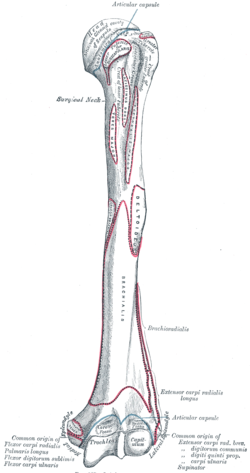
The humerus is a long bone in the arm that runs from the shoulder to the elbow. It connects the scapula and the two bones of the lower arm, the radius and ulna, and consists of three sections. The humeral upper extremity consists of a rounded head, a narrow neck, and two short processes. The body is cylindrical in its upper portion, and more prismatic below. The lower extremity consists of 2 epicondyles, 2 processes, and 3 fossae. As well as its true anatomical neck, the constriction below the greater and lesser tubercles of the humerus is referred to as its surgical neck due to its tendency to fracture, thus often becoming the focus of surgeons.
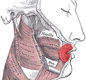
In human anatomy, the orbicularis oris muscle is a complex of muscles in the lips that encircles the mouth. It is not a true sphincter, as was once thought, as it is actually composed of four independent quadrants that interlace and give only an appearance of circularity.
The biceps femoris is a muscle of the thigh located to the posterior, or back. As its name implies, it consists of two heads; the long head is considered part of the hamstring muscle group, while the short head is sometimes excluded from this characterization, as it only causes knee flexion and is activated by a separate nerve.

The vastus medialis is an extensor muscle located medially in the thigh that extends the knee. The vastus medialis is part of the quadriceps muscle group.
The pronator teres is a muscle that, along with the pronator quadratus, serves to pronate the forearm. It has two origins, at the medial humeral supracondylar ridge and the ulnar tuberosity, and inserts near the middle of the radius.

The plantaris is one of the superficial muscles of the superficial posterior compartment of the leg, one of the fascial compartments of the leg.
The linea aspera is a ridge of roughened surface on the posterior surface of the shaft of the femur. It is the site of attachments of muscles and the intermuscular septum.
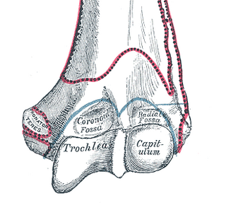
In the human arm, the humeral trochlea is the medial portion of the articular surface of the elbow joint which articulates with the trochlear notch on the ulna in the forearm.
In human anatomy, the body of femur is the almost cylindrical, long part of the femur. It is a little broader above than in the center, broadest and somewhat flattened from before backward below. It is slightly arched, so as to be convex in front, and concave behind, where it is strengthened by a prominent longitudinal ridge, the linea aspera.

The medial condyle is one of the two projections on the lower extremity of femur, the other being the lateral condyle.
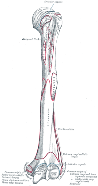
The lateral supracondylar ridge is a prominent, rough margin on the lower part of the lateral border of the humerus. It presents an anterior lip for the origin of forearm extensors, including the brachioradialis muscle above, and the extensor carpi radialis longus muscle below. It also presents a posterior lip for the triceps brachii, and an intermediate ridge for the attachment of the lateral intermuscular septum.

The medial epicondyle of the humerus is an epicondyle of the humerus bone of the upper arm in humans. It is larger and more prominent than the lateral epicondyle and is directed slightly more posteriorly in the anatomical position. In birds, where the arm is somewhat rotated compared to other tetrapods, it is called the ventral epicondyle of the humerus. In comparative anatomy, the more neutral term entepicondyle is used.
The brachial fascia is continuous with that covering the deltoideus and the pectoralis major muscle, by means of which it is attached, above, to the clavicle, acromion, and spine of the scapula; it forms a thin, loose, membranous sheath for the muscles of the arm, and sends septa between them; it is composed of fibers disposed in a circular or spiral direction, and connected together by vertical and oblique fibers.

The fascial compartments of arm refers to the specific anatomical term of the compartments within the upper segment of the upper limb of the body. The upper limb is divided into two segments, the arm and the forearm. Each of these segments is further divided into two compartments which are formed by deep fascia – tough connective tissue septa (walls). Each compartment encloses specific muscles and nerves.

In human anatomy, the adductor hiatus also known as hiatus magnus is a hiatus (gap) between the adductor magnus muscle and the femur that allows the passage of the femoral vessels from the anterior thigh to the posterior thigh and then the popliteal fossa. It is the termination of the adductor canal and lies about 8–13.5 cm (3.1–5.3 in) superior to the adductor tubercle.
Supracondylar ridge may refer to:

Struthers' ligament is a feature of human anatomy consisting of a band of connective tissue at the medial aspect of the distal humerus. It courses from the supracondylar process of the humerus to the medial humeral epicondyle. It is not a constant ligament, and can be acquired or congenital. The structure was highlighted by John Struthers, who discussed the feature's evolutionary significance with Charles Darwin. Struthers originally reported that the ligament usually arose at a position 3.2 to 6.4 cm from the medial condyle, being 1.2 to 1.9 cm in length, and nearer to the anterior than the medial border of the humerus.
The vastus muscles are three of the four muscles that make up the quadriceps femoris muscle of the thigh. The three muscles are the vastus intermedius, the vastus lateralis, and the vastus medialis located in the middle, on the outside, and inside of the thigh, respectively. The fourth muscle is the rectus femoris muscle a large fleshy muscle which covers the front and sides of the femur.

The supracondylar process of the humerus is a variant bony projection on the anteromedial aspect of the upper arm bone (humerus), about 5–6 cm above the medial epicondyle. It is directed downward, forward and medially pointing to the medial epicondyle. A fibrous band, Struthers ligament, may connect this process to the medial epicondyle. This variation has a prevalence of 0.68% and is significantly more common in women than in men.