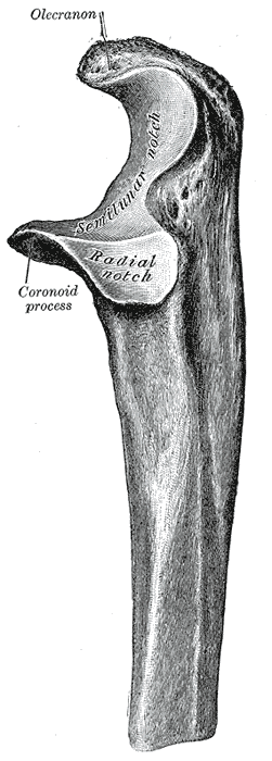
Coronoid process of the ulna

Last updated
Coronoid process of the ulna
Gray212.png
Upper extremity of left ulna. Lateral aspect.
Details
Identifiers
Latin Processus coronoideus ulnae
TA98 A02.4.06.003
TA2 1232
FMA 23616
Anatomical terms of bone

The coronoid process of the ulna is a triangular process projecting forward from the anterior proximal portion of the ulna.

Contents

Structure

Its base is continuous with the body of the bone, and of considerable strength.

Its apex is pointed, slightly curved upward, and in flexion of the forearm is received into the coronoid fossa of the humerus.

Its upper surface is smooth, convex, and forms the lower part of the semilunar notch.

Its antero-inferior surface is concave, and marked by a rough impression for the insertion of the brachialis muscle. At the junction of this surface with the front of the body is a rough eminence, the tuberosity of the ulna, which gives insertion to a part of the brachialis; to the lateral border of this tuberosity the oblique cord is attached.

Its lateral surface presents a narrow, oblong, articular depression, the radial notch.

Its medial surface, by its prominent, free margin, serves for the attachment of part of the ulnar collateral ligament. At the front part of this surface is a small rounded eminence for the origin of one head of the flexor digitorum superficialis muscle; behind the eminence is a depression for part of the origin of the flexor digitorum profundus muscle; descending from the eminence is a ridge which gives origin to one head of the pronator teres muscle.

Frequently, the flexor pollicis longus muscle arises from the lower part of the coronoid process by a rounded bundle of muscular fibers.

Function

The coronoid process stabilises the elbow joint and prevents hyperflexion. [1] [2]

Clinical significance

The coronoid process can be fractured from its anteromedial facet. [1] [3]

Additional images

Related Research Articles

<span class="mw-page-title-main">Ulna</span> Medial bone from forearm

The ulna or ulnal bone is a long bone found in the forearm that stretches from the elbow to the wrist, and when in anatomical position, is found on the medial side of the forearm. That is, the ulna is on the same side of the forearm as the little finger. It runs parallel to the radius, the other long bone in the forearm. The ulna is longer and the radius is shorter, but the radius is thicker and the ulna is thinner. Therefore, the ulna is considered to be the smaller bone of the two bones in the lower arm. The corresponding bone in the lower leg is the fibula.

<span class="mw-page-title-main">Humerus</span> Long bone of the upper arm

The humerus is a long bone in the arm that runs from the shoulder to the elbow. It connects the scapula and the two bones of the lower arm, the radius and ulna, and consists of three sections. The humeral upper extremity consists of a rounded head, a narrow neck, and two short processes. The body is cylindrical in its upper portion, and more prismatic below. The lower extremity consists of 2 epicondyles, 2 processes, and 3 fossae. As well as its true anatomical neck, the constriction below the greater and lesser tubercles of the humerus is referred to as its surgical neck due to its tendency to fracture, thus often becoming the focus of surgeons.

<span class="mw-page-title-main">Median nerve</span> Nerve of the upper limb

The median nerve is a nerve in humans and other animals in the upper limb. It is one of the five main nerves originating from the brachial plexus.

<span class="mw-page-title-main">Brachialis muscle</span> Flexor muscle in the upper arm

The brachialis is a muscle in the upper arm that flexes the elbow. It lies beneath the biceps brachii, and makes up part of the floor of the region known as the cubital fossa. It originates from the anterior aspect of the distal humerus; it inserts onto the tuberosity of the ulna. It is innervated by the musculocutaneous nerve, and commonly also receives additional innervation from the radial nerve. The brachialis is the prime mover of elbow flexion generating about 50% more power than the biceps.

<span class="mw-page-title-main">Tibia</span> Leg bone in vertebrates

The tibia, also known as the shinbone or shankbone, is the larger, stronger, and anterior (frontal) of the two bones in the leg below the knee in vertebrates ; it connects the knee with the ankle. The tibia is found on the medial side of the leg next to the fibula and closer to the median plane. The tibia is connected to the fibula by the interosseous membrane of leg, forming a type of fibrous joint called a syndesmosis with very little movement. The tibia is named for the flute tibia. It is the second largest bone in the human body, after the femur. The leg bones are the strongest long bones as they support the rest of the body.

<span class="mw-page-title-main">Radius (bone)</span> One of the two long bones of the forearm

The radius or radial bone is one of the two large bones of the forearm, the other being the ulna. It extends from the lateral side of the elbow to the thumb side of the wrist and runs parallel to the ulna. The ulna is longer than the radius, but the radius is thicker. The radius is a long bone, prism-shaped and slightly curved longitudinally.

<span class="mw-page-title-main">Upper limb</span> Consists of the arm, forearm, and hand

The upper limbs or upper extremities are the forelimbs of an upright-postured tetrapod vertebrate, extending from the scapulae and clavicles down to and including the digits, including all the musculatures and ligaments involved with the shoulder, elbow, wrist and knuckle joints. In humans, each upper limb is divided into the arm, forearm and hand, and is primarily used for climbing, lifting and manipulating objects.

<span class="mw-page-title-main">Musculocutaneous nerve</span> Nerve in the arm

The musculocutaneous nerve is a mixed branch of the lateral cord of the brachial plexus derived from cervical spinal nerves C5-C7. It arises opposite the lower border of the pectoralis major. It provides motor innervation to the muscles of the anterior compartment of the arm: the coracobrachialis, biceps brachii, and brachialis. It provides sensory innervation to the lateral forearm. It courses through the anterior part of the arm, terminating 2 cm above elbow; after passing the lateral edge of the tendon of biceps brachii it is becomes known as the lateral cutaneous nerve of the forearm.

<span class="mw-page-title-main">Ulnar artery</span> Artery of the forearm

The ulnar artery is the main blood vessel, with oxygenated blood, of the medial aspects of the forearm. It arises from the brachial artery and terminates in the superficial palmar arch, which joins with the superficial branch of the radial artery. It is palpable on the anterior and medial aspect of the wrist.

The flexor pollicis longus is a muscle in the forearm and hand that flexes the thumb. It lies in the same plane as the flexor digitorum profundus. This muscle is unique to humans, being either rudimentary or absent in other primates. A meta-analysis indicated accessory flexor pollicis longus is present in around 48% of the population.

The pronator teres is a muscle that, along with the pronator quadratus, serves to pronate the forearm. It has two origins, at the medial humeral supracondylar ridge and the ulnar tuberosity, and inserts near the middle of the radius.

<span class="mw-page-title-main">Supinator muscle</span> Muscle of the forearm in humans

In human anatomy, the supinator is a broad muscle in the posterior compartment of the forearm, curved around the upper third of the radius. Its function is to supinate the forearm.

<span class="mw-page-title-main">Anterior interosseous artery</span>

The anterior interosseous artery is an artery in the forearm. It is a branch of the common interosseous artery.

<span class="mw-page-title-main">Trochlea of humerus</span> Articular surface of the elbow joint which articulates with the trochlear notch of the ulna

In the human arm, the humeral trochlea is the medial portion of the articular surface of the elbow joint which articulates with the trochlear notch on the ulna in the forearm.

<span class="mw-page-title-main">Medial epicondyle of the humerus</span> Rounded eminence on the medial side of the humerus

The medial epicondyle of the humerus is an epicondyle of the humerus bone of the upper arm in humans. It is larger and more prominent than the lateral epicondyle and is directed slightly more posteriorly in the anatomical position. In birds, where the arm is somewhat rotated compared to other tetrapods, it is called the ventral epicondyle of the humerus. In comparative anatomy, the more neutral term entepicondyle is used.

<span class="mw-page-title-main">Anterior interosseous nerve</span>

The anterior interosseous nerve is a branch of the median nerve that supplies the deep muscles on the anterior of the forearm, except the ulnar (medial) half of the flexor digitorum profundus. Its nerve roots come from C8 and T1.

<span class="mw-page-title-main">Tuberosity of the ulna</span>

The tuberosity of the ulna is a rough eminence on the proximal end of the ulna. It occurs at the junction of the antero-inferior surface of the coronoid process with the front of the body. It provides an insertion point to a tendon of the brachialis.

<span class="mw-page-title-main">Elbow</span> Joint between the upper and lower parts of the arm

The elbow is the region between the upper arm and the forearm that surrounds the elbow joint. The elbow includes prominent landmarks such as the olecranon, the cubital fossa, and the lateral and the medial epicondyles of the humerus. The elbow joint is a hinge joint between the arm and the forearm; more specifically between the humerus in the upper arm and the radius and ulna in the forearm which allows the forearm and hand to be moved towards and away from the body. The term elbow is specifically used for humans and other primates, and in other vertebrates forelimb plus joint is used.

<span class="mw-page-title-main">Pronator teres syndrome</span> Medical condition

Pronator teres syndrome is a compression neuropathy of the median nerve at the elbow. It is rare compared to compression at the wrist or isolated injury of the anterior interosseous branch of the median nerve.

<span class="mw-page-title-main">Median nerve palsy</span> Medical condition

Injuries to the arm, forearm or wrist area can lead to various nerve disorders. One such disorder is median nerve palsy. The median nerve controls the majority of the muscles in the forearm. It controls abduction of the thumb, flexion of hand at wrist, flexion of digital phalanx of the fingers, is the sensory nerve for the first three fingers, etc. Because of this major role of the median nerve, it is also called the eye of the hand. If the median nerve is damaged, the ability to abduct and oppose the thumb may be lost due to paralysis of the thenar muscles. Various other symptoms can occur which may be repaired through surgery and tendon transfers. Tendon transfers have been very successful in restoring motor function and improving functional outcomes in patients with median nerve palsy.

References

PD-icon.svgThis article incorporates text in the public domain from page 214 of the 20th edition of Gray's Anatomy (1918)

  1. 1 2 Doornberg, Job N.; de Jong, Inge M.; Lindenhovius, Anneluuk L. C.; Ring, David (2007-09-01). "The anteromedial facet of the coronoid process of the ulna". Journal of Shoulder and Elbow Surgery. 16 (5): 667–670. doi:10.1016/j.jse.2007.03.013. ISSN   1058-2746. PMID   17512221.
  2. Ni, Qubo; Yang, Xu; Pan, Zhengqi; Wang, Jianping (2020-05-14). "The pronator teres and the flexor carpi radialis interval approach for operative fixation of ulna coronoid process fractures". Orthopaedics & Traumatology: Surgery & Research. 107 (2): 102610. doi: 10.1016/j.otsr.2020.04.004 . ISSN   1877-0568. PMID   32418740.
  3. Sanchez-Sotelo, Joaquin; O’Driscoll, Shawn W.; Morrey, Bernard F. (2005-01-01). "Medial oblique compression fracture of the coronoid process of the ulna". Journal of Shoulder and Elbow Surgery. 14 (1): 60–64. doi:10.1016/j.jse.2004.04.012. ISSN   1058-2746. PMID   15723014.