 Front of the left forearm. Deep muscles.
 Cross-section through the middle of the forearm.
 Posterior surface of the forearm. Deep muscles.
 Transverse section across distal ends of radius and ulna.
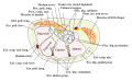 Transverse section across the wrist and digits.
 The mucous sheaths of the tendons on the front of the wrist and digits.
 Ulnar and radial arteries. Deep view.
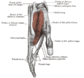 Tendons of forefinger and vincula tendina. (Flexor digitorum profundus labeled at bottom right.)
 Flexor digitorum profundus muscle
 Flexor digitorum profundus muscle
 Flexor digitorum profundus muscle
 Flexor digitorum profundus muscle
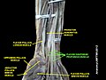 Flexor digitorum profundus muscle
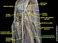 Flexor digitorum profundus muscle
 Flexor digitorum profundus muscle
 Flexor digitorum profundus muscle
 Flexor digitorum profundus muscle
| Flexor digitorum profundus muscle | |
|---|---|
 Ventral view of the deep muscles of the left forearm. FDP is shown in blue. | |
| Details | |
| Origin | Upper 3/4 of the anterior and medial surfaces of the body of the ulna, interosseous membrane and deep fascia of the forearm |
| Insertion | Base of the distal phalanges of the fingers |
| Artery | Anterior interosseous artery |
| Nerve | Lateral part of the muscle by median (anterior interosseous), medial (ulnar) part of the muscle by muscular branches of ulnar |
| Actions | Flex hand and both interphalangeal joints |
| Antagonist | Extensor digitorum muscle |
| Identifiers | |
| Latin | musculus flexor digitorum profundus |
| TA98 | A04.6.02.036 |
| TA2 | 2491 |
| FMA | 38478 |
| Anatomical terms of muscle | |
The flexor digitorum profundus or flexor digitorum communis profundus [1] is a muscle in the forearm of humans that flexes the fingers (also known as digits). It is considered an extrinsic hand muscle because it acts on the hand while its muscle belly is located in the forearm.
Contents
- Structure
- Nerve supply
- Variation
- Function
- Other animals
- Additional images
- See also
- Notes
- References
- External links
Together the flexor pollicis longus, pronator quadratus, and flexor digitorum profundus form the deep layer of ventral forearm muscles. [2] The muscle is named from Latin 'deep bender of the fingers'.


