Subscapularis muscle

Last updated

Subscapularis muscle
Subscapularis muscle frontal.png
Subscapularis muscle (in red). Ribs are shown as semi-transparent.
1119 Muscles that Move the Humerus c.png
The subscapularis is difficult to see from the front (labeled middle right)
Details
Origin Subscapular fossa
Insertion Lesser tubercle of humerus
Artery Subscapular artery
Nerve Upper subscapular nerve, lower subscapular nerve (C5, C6)
Actions Internally rotates and adducts humerus; stabilizes shoulder
Identifiers
Latin musculus subscapularis
TA98 A04.6.02.012
TA2 2460
FMA 13413
Anatomical terms of muscle

The subscapularis is a large triangular muscle which fills the subscapular fossa and inserts into the lesser tubercle of the humerus and the front of the capsule of the shoulder-joint.

Contents

Structure

The subscapularis is covered by a dense fascia which attaches to the scapula at the margins of the subscapularis' attachment (origin) on the scapula. [1]

The muscle's fibers pass laterally from its origin before coalescing into a tendon of insertion.[ citation needed ] The tendon intermingles with the glenohumeral (shoulder) joint capsule. [1]

A bursa (which communicates with the cavity of the shoulder joint [1] [2] via an aperture in the joint capsule [2] ) intervenes between the tendon and a bare area at the lateral angle of the scapula [1] /the neck of the scapula. [2] The subscapularis (supraserratus) bursa separates the subscapularis is from the serratus anterior. [2]

Origin

It arises from its medial two-thirds of the costal surface of the scapula, the intermuscular septa (which create ridges upon the scapula), [1] and the lower two-thirds of the groove on the axillary border (subscapular fossa) of the scapula.[ citation needed ]

Some fibers arise from tendinous laminae, which intersect the muscle and are attached to ridges on the bone; others from an aponeurosis, which separates the muscle from the teres major and the long head of the triceps brachii.[ citation needed ]

Insertion

It inserts onto the lesser tubercle of the humerus [1] and the anterior part of the shoulder-joint capsule. Tendinous fibers extend to the greater tubercle with insertions into the bicipital groove.[ citation needed ]

Innervation

The subscapularis is supplied by the upper and lower subscapular nerves (C5-C6), branches of the posterior cord of the brachial plexus. [1]

Actions/movements

The subscapularis medially (internally) rotates the humerus (acting here as a prime mover) [1] and adducts it. When the arm is raised, it draws the humerus forward and downward.[ citation needed ]

Function

The subscapularis stabilises the shoulder joint by contributing to the fixation of the proximal humerus during movements of the elbow, wrist, and hand. [1] It is a powerful defense to the front of the shoulder-joint, preventing displacement of the head of the humerus.[ citation needed ]

Clinical significance

Examination

It is difficult to isolate the action of the subscapularis from other medial rotators of the shoulder joint; there is no satisfactory test for this muscle. [1] The Gerber Lift-off test is the established clinical test for examination of the subscapularis. [3] The bear hug test (internal rotation while palm is held on opposite shoulder and elbow is held in a position of maximal anterior translation) for subscapularis muscle tears has high sensitivity. Positive bear-hug and belly press tests indicate significant tearing of subscapularis. [4]

Imaging

MRI. Partial rupture of the cranial subscapularis tendon at the insertion site MRI. Partial rupture of the cranial subscapularis tendon at the insertion site..jpg
MRI. Partial rupture of the cranial subscapularis tendon at the insertion site

There is no singularly imaging device or technique for a satisfying and complete subscapularis examination, but rather the combination of the sagittal oblique MRI / short-axis US and axial MRI / long-axis US planes seems to generate useful results. Additionally, lesser tuberosity bony changes have been associated with subscapularis tendon tears. Findings with cysts seem to be more specific and combined findings with cortical irregularities more sensitive. [5]

Another fact typically for the subscapularis muscle is the fatty infiltration of the superior portions, while sparing the inferior portions.

Since the long biceps tendon absents itself from the shoulder joint through the rotator cuff interval, it is easily possible to distinguish between the supraspinatus and the subscapularis tendon. Those two tendons build the interval sling.

Ultrasonography

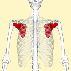
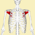
Mack et al. developed an ultrasonographic procedure with which it is possible to explore almost the complete rotator cuff within six steps. It unveils clearly the whole area from the subedge of the subscapularis tendon until the intersection between the infraspinatus tendon and musculus teres minor. One of six steps does focus on the subscapularis tendon. In the first instance the examinator guides the applicator to the proximal humerus as perpendicularly as possible to the sulcus intertubercularis. Gliding now medially shows the insertion of the subscapularis tendon. [6]

Longitudinal plane of the musculus subscapularis and its tendon

The subscapularis tendon lies approximately 3 to 5 cm under the surface. Quite deep for ultrasonography, and therefore displaying through a highly penetrative 5 MHz linear applicator is worth a try. And it really turned out to ease a detailed examination of the muscle which just abuts to the scapula. However, the tendon of primary interest does not get mapped as closely as desired. As anatomical analysis showed, it is only by external rotation possible to see the ventral part of the joint socket and its labrum. While at the neutral position the tuberculum minus occludes the view. Summing up it is through an external arm rotation and a medially applied 5 MHz sector sonic head possible to display the ventral part of the joint socket and its labrum with notedly lower echogenicity. [7]

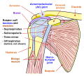
The following sectional planes are defined for the sonographic examination of the different shoulder joint structures: [8]

Ventral transversalVentral sagittal medialVentral sagittal lateralLateral coronalLateral transversal/sagittal:Dorsal transversalDorsal sagittal
subscapularis muscle (longitudinal)subscapularis muscle (transversal)Intertub. sulcus with long head of biceps brachii (longitudinal)supraspinatus muscle (longitudinal)supraspinatus muscle (transversal)infraspinatus muscle (longitudinal)supraspinatus muscle (transversal)
Intertubercular sulcus with long head of biceps brachii (transversal)Hill-Sachs-Lesio

Tissue harmonic imaging

Primarily in abdominal imaging, tissue harmonic imaging (THI) gets more and more valued and used additionally to conventional ultrasonography.

THI involves the use of harmonic frequencies that originate within the tissue as a result of nonlinear wave front propagation and are not present in the incident beam. These harmonic signals may arise differently at anatomic sites with similar impedances and thus lead to higher contrast resolution." Along with higher contrast resolution it has an elevated signal-to-noise ratio and significantly reduced inter- and intraobserver variability compared with conventional US. Additionally it is possible to nearly eliminate ordinary US artifacts, i.e. side-lobe, near-field artifacts, reverberation artifacts. As aforementioned THI has already led to enhanced abdominal, breast, vascular and cardiac sonography.

For musculo-skeletal aspects THI has not been used that much, although this method features some useful potential. For example, for the still tricky discrimination between the presence of a hypoechoic defect and/or loss of the outer tendon convexity/non-visualization of the tendon, that is between partial- and full-thickness rotator cuff tears.

In comparison to a checking MR Arthrography Strobel K. et al. has arrived at the conclusion that through THI it is possible to achieve a generally improved visibility of joint and tendon surfaces, especially superior for subscapularis tendon abnormalities. [9]

Additional images

Related Research Articles

<span class="mw-page-title-main">Scapula</span> Bone that connects the humerus (upper arm bone) with the clavicle (collar bone)

The scapula, also known as the shoulder blade, is the bone that connects the humerus with the clavicle. Like their connected bones, the scapulae are paired, with each scapula on either side of the body being roughly a mirror image of the other. The name derives from the Classical Latin word for trowel or small shovel, which it was thought to resemble.

<span class="mw-page-title-main">Humerus</span> Long bone of the upper arm

The humerus is a long bone in the arm that runs from the shoulder to the elbow. It connects the scapula and the two bones of the lower arm, the radius and ulna, and consists of three sections. The humeral upper extremity consists of a rounded head, a narrow neck, and two short processes. The body is cylindrical in its upper portion, and more prismatic below. The lower extremity consists of 2 epicondyles, 2 processes, and 3 fossae. As well as its true anatomical neck, the constriction below the greater and lesser tubercles of the humerus is referred to as its surgical neck due to its tendency to fracture, thus often becoming the focus of surgeons.

<span class="mw-page-title-main">Rotator cuff</span> Group of muscles

The rotator cuff is a group of muscles and their tendons that act to stabilize the human shoulder and allow for its extensive range of motion. Of the seven scapulohumeral muscles, four make up the rotator cuff. The four muscles are:

<span class="mw-page-title-main">Biceps</span> Muscle on the front of the upper arm

The biceps or biceps brachii are a large muscle that lies on the front of the upper arm between the shoulder and the elbow. Both heads of the muscle arise on the scapula and join to form a single muscle belly which is attached to the upper forearm. While the biceps crosses both the shoulder and elbow joints, its main function is at the elbow where it flexes the forearm and supinates the forearm. Both these movements are used when opening a bottle with a corkscrew: first biceps screws in the cork (supination), then it pulls the cork out (flexion).

<span class="mw-page-title-main">Shoulder problem</span> Medical condition

Shoulder problems including pain, are one of the more common reasons for physician visits for musculoskeletal symptoms. The shoulder is the most movable joint in the body. However, it is an unstable joint because of the range of motion allowed. This instability increases the likelihood of joint injury, often leading to a degenerative process in which tissues break down and no longer function well.

<span class="mw-page-title-main">Shoulder</span> Part of the body

The human shoulder is made up of three bones: the clavicle (collarbone), the scapula, and the humerus as well as associated muscles, ligaments and tendons.

<span class="mw-page-title-main">Deltoid muscle</span> Shoulder muscle

The deltoid muscle is the muscle forming the rounded contour of the human shoulder. It is also known as the 'common shoulder muscle', particularly in other animals such as the domestic cat. Anatomically, the deltoid muscle appears to be made up of three distinct sets of muscle fibers, namely the

  1. anterior or clavicular part
  2. posterior or scapular part
  3. intermediate or acromial part
<span class="mw-page-title-main">Upper limb</span> Consists of the arm, forearm, and hand

The upper limbs or upper extremities are the forelimbs of an upright-postured tetrapod vertebrate, extending from the scapulae and clavicles down to and including the digits, including all the musculatures and ligaments involved with the shoulder, elbow, wrist and knuckle joints. In humans, each upper limb is divided into the arm, forearm and hand, and is primarily used for climbing, lifting and manipulating objects.

<span class="mw-page-title-main">Teres minor muscle</span> Muscle of the rotator cuff

The teres minor is a narrow, elongated muscle of the rotator cuff. The muscle originates from the lateral border and adjacent posterior surface of the corresponding right or left scapula and inserts at both the greater tubercle of the humerus and the posterior surface of the joint capsule.

<span class="mw-page-title-main">Triceps</span> Muscle on the back of the upper arm

The triceps, or triceps brachii, is a large muscle on the back of the upper limb of many vertebrates. It consists of 3 parts: the medial, lateral, and long head. It is the muscle principally responsible for extension of the elbow joint.

<span class="mw-page-title-main">Supraspinatus muscle</span> Muscle of the upper back

The supraspinatus is a relatively small muscle of the upper back that runs from the supraspinous fossa superior portion of the scapula to the greater tubercle of the humerus. It is one of the four rotator cuff muscles and also abducts the arm at the shoulder. The spine of the scapula separates the supraspinatus muscle from the infraspinatus muscle, which originates below the spine.

<span class="mw-page-title-main">Infraspinatus muscle</span> Main external rotator of the shoulder

In human anatomy, the infraspinatus muscle is a thick triangular muscle, which occupies the chief part of the infraspinatous fossa. As one of the four muscles of the rotator cuff, the main function of the infraspinatus is to externally rotate the humerus and stabilize the shoulder joint.

<span class="mw-page-title-main">Shoulder joint</span> Synovial ball and socket joint in the shoulder

The shoulder joint is structurally classified as a synovial ball-and-socket joint and functionally as a diarthrosis and multiaxial joint. It involves an articulation between the glenoid fossa of the scapula and the head of the humerus. Due to the very loose joint capsule that gives a limited interface of the humerus and scapula, it is the most mobile joint of the human body.

<span class="mw-page-title-main">Teres major muscle</span> Muscle of the upper limb

The teres major muscle is a muscle of the upper limb. It attaches to the scapula and the humerus and is one of the seven scapulohumeral muscles. It is a thick but somewhat flattened muscle.

<span class="mw-page-title-main">Glenoid fossa</span> Part of the shoulder

The glenoid fossa of the scapula or the glenoid cavity is a bone part of the shoulder. The word glenoid is pronounced or and is from Greek: gléne, "socket", reflecting the shoulder joint's ball-and-socket form. It is a shallow, pyriform articular surface, which is located on the lateral angle of the scapula. It is directed laterally and forward and articulates with the head of the humerus; it is broader below than above and its vertical diameter is the longest.

<span class="mw-page-title-main">Lesser tubercle</span> Bony projection on the proximal end of the humerus

The lesser tubercle of the humerus, although smaller, is more prominent than the greater tubercle: it is situated in front, and is directed medially and anteriorly.

<span class="mw-page-title-main">Greater tubercle</span> Bony projection on the proximal end of the humerus

The greater tubercle of the humerus is the outward part the upper end of that bone, adjacent to the large rounded prominence of the humerus head. It provides attachment points for the supraspinatus, infraspinatus, and teres minor muscles, three of the four muscles of the rotator cuff, a muscle group that stabilizes the shoulder joint. In doing so the tubercle acts as a location for the transfer of forces from the rotator cuff muscles to the humerus.

<span class="mw-page-title-main">Spine of scapula</span> Bony plate on the scapula

The spine of the scapula or scapular spine is a prominent plate of bone, which crosses obliquely the medial four-fifths of the scapula at its upper part, and separates the supra- from the infraspinatous fossa.

<span class="mw-page-title-main">Shoulder impingement syndrome</span> Medical condition

Shoulder impingement syndrome is a syndrome involving tendonitis of the rotator cuff muscles as they pass through the subacromial space, the passage beneath the acromion. It is particularly associated with tendonitis of the supraspinatus muscle. This can result in pain, weakness, and loss of movement at the shoulder.

<span class="mw-page-title-main">Oblique ridges of scapula</span>

The oblique ridges cross the subscapular fossa from superomedial to inferiolateral. These ridges are formed by intramuscular tendons of the subscapularis muscle.

References

PD-icon.svgThis article incorporates text in the public domain from page 440 of the 20th edition of Gray's Anatomy (1918)

  1. 1 2 3 4 5 6 7 8 9 10 Sinnatamby, Chummy (2011). Last's Anatomy (12th ed.). pp. 44–45. ISBN   978-0-7295-3752-0.
  2. 1 2 3 4 Milano, Giuseppe and Grasso, Andrea, Shoulder Arthroscopy: Principles and Practice Archived 27 April 2021 at the Wayback Machine , Springer Science & Business Media, Dec 16, 2013. ISBN   9781447154273. Accessed 2016-11-07.
  3. Simons, Stephen; Dixon, J Bryan (3 April 2017). "Physical examination of the shoulder" . UpToDate. Archived from the original on 20 April 2017. Retrieved 19 April 2017.
  4. Barth, Johannes R.H.; Burkhart, Stephen S.; De Beer, Joe F. (October 2006). "The Bear-Hug Test: A New and Sensitive Test for Diagnosing a Subscapularis Tear". Arthroscopy: The Journal of Arthroscopic and Related Surgery. 22 (10): 1076–1084. doi:10.1016/j.arthro.2006.05.005. PMID   17027405.
  5. Morag, Yoav; Jamadar, David A.; Miller, Bruce; Dong, Qian; Jacobson, Jon A. (2009). "The subscapularis: Anatomy, injury, and imaging". Skeletal Radiology. 40 (3): 255–69. doi:10.1007/s00256-009-0845-0. PMID   20033149. S2CID   13833846.
  6. Mack, L A; Matsen, F A; Kilcoyne, R F; Davies, P K; Sickler, M E (1985). "US evaluation of the rotator cuff". Radiology. 157 (1): 205–9. doi:10.1148/radiology.157.1.3898216. PMID   3898216.
  7. Katthagen BD. et al.. Schultersonographie. Stuttgart. ISBN   3-13-719401-6 [ page needed ]
  8. Thelen M. et al.. Radiologische Diagnostik der Verletzungen von Knochen und Gelenken. Stuttgart [etc.]. Georg Thieme. 1993. ISBN   3-13-778701-7 [ page needed ]
  9. Strobel, Klaus; Zanetti, Marco; Nagy, Ladislav; Hodler, Juerg (2004). "Suspected Rotator Cuff Lesions: Tissue Harmonic Imaging versus Conventional US of the Shoulder1". Radiology. 230 (1): 243–9. doi:10.1148/radiol.2301021517. PMID   14631052.