 Bones of left forearm. Posterior aspect.
 Tendons of forefinger and vincula tendina.
 Cross-section through the middle of the forearm.
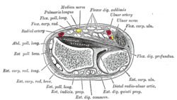 Transverse section across distal ends of radius and ulna.
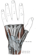 The mucous sheaths of the tendons on the back of the wrist.
 Extensor digitorum communis muscle
 Extensor digitorum muscles
 Extensor digitorum muscle
 Extensor digitorum muscle
 Extensor digitorum muscle
 Extensor digitorum muscle
 Extensor digitorum muscle
 Extensor digitorum muscle
 Extensor digitorum muscle
 Extensor digitorum muscle
 Muscles of hand. Posterior view.
 Muscles of hand. Posterior view.
| Extensor digitorum muscle | |
|---|---|
 Posterior surface of the forearm. Superficial muscles. Extensor digitorum muscle is labeled in purple. | |
 Transverse section across the wrist and digits. (Ext. dig. communis labeled at bottom center.) | |
| Details | |
| Origin | Lateral epicondyle (common extensor tendon) |
| Insertion | Extensor expansion of middle and distal phalanges of the 2nd, 3rd, 4th, and 5th fingers [1] |
| Artery | Posterior interosseous artery |
| Nerve | Posterior interosseous nerve |
| Actions | Extension of hand, wrist and fingers |
| Antagonist | Flexor digitorum superficialis muscle, flexor digitorum profundus muscle |
| Identifiers | |
| Latin | musculus extensor digitorum |
| TA98 | A04.6.02.042 |
| TA2 | 2500 |
| FMA | 38500 |
| Anatomical terms of muscle | |
The extensor digitorum muscle (also known as extensor digitorum communis) [2] is a muscle of the posterior forearm present in humans and other animals. It extends the medial four digits of the hand. Extensor digitorum is innervated by the posterior interosseous nerve, which is a branch of the radial nerve. [3]


