Juncturae tendinum

Last updated
Juncturae tendinum
Sobo 1909 289.png
Juncturae tendinum are seen between the tendons of the extensor digitorum communis.
Details
Identifiers
Latin Juncturae tendinum
Anatomical terms of muscle

In human anatomy, juncturae tendinum or connexus intertendinei refers to the connective tissues that link the tendons of the extensor digitorum communis, and sometimes, to the tendon of the extensor digiti minimi. [1] Juncturae tendinum are located on the dorsal aspect of the hand in the first, second and third inter-metacarpal spaces proximal to the metacarpophalangeal joint.

Connective tissue type of biological tissue

Connective tissue (CT) is one of the four basic types of animal tissue, along with epithelial tissue, muscle tissue, and nervous tissue. It develops from the mesoderm. Connective tissue is found in between other tissues everywhere in the body, including the nervous system. In the central nervous system, the three outer membranes that envelop the brain and spinal cord are composed of connective tissue. They support and protect the body. All connective tissue consists of three main components: fibers, ground substance and cells. Not all authorities include blood or lymph as connective tissue because they lack the fiber component. All are immersed in the body water.

Extensor digitorum muscle

The extensor digitorum muscle is a muscle of the posterior forearm present in humans and other animals. It extends the medial four digits of the hand. Extensor digitorum is innervated by the posterior interosseous nerve, which is a branch of the radial nerve.

Extensor digiti minimi muscle muscle of the forearm

The extensor digiti minimi is a slender muscle of the forearm, placed on the ulnar side of the extensor digitorum communis, with which it is generally connected.

Contents

Structure

Juncturae tendinum are narrow bands of connective tissues that extend between the tendons of the extensor digitorum communis and the extensor digiti minimi. [1] [2] It is classified into three distinct types (Type 1, 2 and 3) depending on morphology. [3]

Morphology (biology) In biology, the form and structure of organisms

Morphology is a branch of biology dealing with the study of the form and structure of organisms and their specific structural features.

Histology

Histological structure of type 1 consists of tight tissue fibers and loose connective tissues. [4] The surrounding loose fibers are rich in blood vessels with pathways for nerves. [4] Tendinous fibers are rarely found and are in single direction. [4] Type 2 juncturae tendinum is also composed of single direction fibers like type 1, but with thicker bundles. [4] Histological structure of the type 3 juncturae is denser and thicker than type 2 juncturae and tendinous fibers are arranged in two layers. Tendinous fibers of the type 3 show crosswise direction with single direction and oblique bundles. [4] No muscular tissue is observed in any type of the juncturae tendinum.

Histology Study of the microscopic anatomy of cells and tissues of plants and animals

Histology, also known as microscopic anatomy or microanatomy, is the branch of biology which studies the microscopic anatomy of biological tissues. Histology is the microscopic counterpart to gross anatomy, which looks at larger structures visible without a microscope. Although one may divide microscopic anatomy into organology, the study of organs, histology, the study of tissues, and cytology, the study of cells, modern usage places these topics under the field of histology. In medicine, histopathology is the branch of histology that includes the microscopic identification and study of diseased tissue. In the field of paleontology, the term paleohistology refers to the histology of fossil organisms.

Variations

Type 1 juncturae is mostly seen between the index and the middle fingers (second inter metacarpal space). [3] [4] [5] Type 2 juncturae is observed mainly between the middle and the ring fingers (third inter-metacarpal space). [2] [3] [4] [5] Type 3 juncturae tendinum is mostly found between the ring and the little finger (fourth inter-metacarpal space). [2] [3] [4] [5]

Normally, the extensor indicis proprius do not receive any juncture tendinum, [3] [6] but it is rarely seen and the mobility of the index finger is compromised. [2] The absence of the tendon of the extensor digitorum communis to the little finger is associated with the thicker type 3r juncturae tendinum in the fourth intermetacarpal space. [3]

Hirai et al. reported type 1 junturae tendinum between the extensor digiti minimi and the extensor digitorum communis to the ring finger in 6% of the specimens. [1]

Functions

Functionalities of juncturae tendinum include coordinating, force distributing, maintaining the space between the common extensor tendons during the extension of different fingers, and stabilizing the metacarpophalangeal joint. [2] [7] [8] Juncturae tendinum also acts as a compensatory slip for the little finger when the tendon of the extensor digitorum comunis to the little finger is absent. [9] However, the presence of juncturae tendinum may make it more difficult to extend each finger independently. [6] [10]

Additional image(s)

Related Research Articles

Wrist part of the arm between the lower arm and the hand

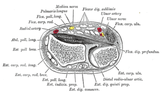
In human anatomy, the wrist is variously defined as 1) the carpus or carpal bones, the complex of eight bones forming the proximal skeletal segment of the hand; (2) the wrist joint or radiocarpal joint, the joint between the radius and the carpus and (3) the anatomical region surrounding the carpus including the distal parts of the bones of the forearm and the proximal parts of the metacarpus or five metacarpal bones and the series of joints between these bones, thus referred to as wrist joints. This region also includes the carpal tunnel, the anatomical snuff box, bracelet lines, the flexor retinaculum, and the extensor retinaculum.

Extensor carpi radialis longus muscle

The extensor carpi radialis longus is one of the five main muscles that control movements at the wrist. This muscle is quite long, starting on the lateral side of the humerus, and attaching to the base of the second metacarpal bone.

Little finger smallest finger of the human hand

The little finger, or pinky finger, also known as the fifth digit, or pinkie, is the most ulnar and smallest finger of the human hand, opposite the thumb, and next to the ring finger.

Palmar interossei muscles

In human anatomy, the palmar or volar interossei are three small, unipennate muscles in the hand that lie between the metacarpal bones and are attached to the index, ring, and little fingers. They are smaller than the dorsal interossei of the hand.

Dorsal interossei of the hand

In human anatomy, the dorsal interossei (DI) are four muscles in the back of the hand that act to abduct (spread) the index, middle, and ring fingers away from hand's midline and assist in flexion at the metacarpophalangeal joints and extension at the interphalangeal joints of the index, middle and ring fingers.

Abductor digiti minimi muscle of hand

In human anatomy, the abductor digiti minimi is a skeletal muscle situated on the ulnar border of the palm of the hand. It forms the ulnar border of the palm and its spindle-like shape defines the hypothenar eminence of the palm together with the skin, connective tissue, and fat surrounding it. Its main function is to pull the little finger away from the other fingers.

Extensor indicis muscle

In human anatomy, the extensor indicis [proprius] is a narrow, elongated skeletal muscle in the deep layer of the dorsal forearm, placed medial to, and parallel with, the extensor pollicis longus. Its tendon goes to the index finger, which it extends.

Sole (foot) bottom part of a foot

The sole is the bottom of the foot.

Vincula tendina

Within each osseo-aponeurotic canal, the tendons of the flexor digitorum superficialis and flexor digitorum profundus are connected to each other, and to the phalanges, by slender, tendinous bands, called vincula tendina.

The posterior compartment of the forearm contains twelve muscles which are chiefly responsible for extension of the wrist and digits, and supination of the forearm. It is separated from the anterior compartment by the interosseous membrane between the radius and ulna.

Palmar plate

In the human hand, palmar or volar plates are found in the metacarpophalangeal (MCP) and interphalangeal (IP) joints where they reinforce the joint capsules, enhance joint stability, and limit hyperextension. The plates of the MCP and IP joints are structurally and functionally similar, except that in the MCP joints they are interconnected by a deep transverse ligament. In the MCP joints, they also indirectly provide stability to the longitudinal palmar arches of the hand. The volar plate of the thumb MCP joint has a transverse longitudinal rectangular shape, shorter than those in the fingers.

Extensor digitorum brevis manus is an extra or accessory muscle on the backside (dorsum) of the hand. It was first described by Albinus in 1758. The muscles lies in the fourth extensor compartment of the wrist, and is relatively rare. It has a prevalence of 4% in the general population according to a meta-analysis. This muscle is commonly misdiagnosed as a ganglion cysta, synovial nodule or cyst.

Mucous sheaths on back of wrist

The mucous sheaths of the tendons on the back of the wrist are protective coverings for tendons in the wrist. Between the dorsal carpal ligament and the bones six compartments are formed for the passage of tendons, each compartment having a separate mucous sheath. One is found in each of the following positions:

  1. on the lateral side of the radial styloid process, for the tendons of the Abductor pollicis longus and Extensor pollicis brevis;
  2. behind the styloid process, for the tendons of the Extensores carpi radialis longus and brevis;
  3. about the middle of the dorsal surface of the radius, for the tendon of the Extensor pollicis longus;
  4. to the medial side of the latter, for the tendons of the Extensor digitorum communis and Extensor indicis proprius;
  5. opposite the interval between the radius and ulna, for the Extensor digiti quinti proprius;
  6. between the head and styloid process of the ulna, for the tendon of the Extensor carpi ulnaris.
Extrinsic extensor muscles of the hand

The extrinsic extensor muscles of the hand are located in the back of the forearm and have long tendons connecting them to bones in the hand, where they exert their action. Extrinsic denotes their location outside the hand. Extensor denotes their action which is to extend, or open flat, joints in the hand. They include the extensor carpi radialis longus (ECRL), extensor carpi radialis brevis (ECRB), extensor digitorum (ED), extensor digiti minimi (EDM), extensor carpi ulnaris (ECU), abductor pollicis longus (APL), extensor pollicis brevis (EPB), extensor pollicis longus (EPL), and extensor indicis (EI).

Extensor medii proprius muscle

The extensor medii proprius is a rare anatomical variant in the extensor compartment of the forearm. The aberrant muscle is analogous to the extensor indicis with the insertion being the middle finger instead of the index finger.

Extensor indicis et medii communis muscle

The extensor indicis et medii communis is a rare anatomical variant in the extensor compartment of forearm. This additional muscle lies in the deep extensor layer next to the extensor indicis proprius and the extensor pollicis longus. The characteristics of this anomalous muscle resemble those of the extensor indicis proprius, with split tendons to the index and the middle finger. This muscle can also be considered as a variation of the aberrant extensor medii proprius.

Extensor pollicis et indicis communis muscle

In human anatomy, the extensor pollicis et indicis communis is an aberrant muscle in the posterior compartment of forearm. It was first described in 1863. The muscle has a prevalence from 0.5% to 4%.

Linburg–Comstock variation is an occasional tendinous connection between the flexor pollicis longus and the flexor digitorum profundus of the index, the middle finger or both. It is found in around 21% of the population. It is an anatomical variation in human, which may be viewed as a pathology if causes symptoms. It was recognised as early as the 1800s, but was first described by Linburg and Comstock in 1979.

References

  1. 1 2 3 Hirai, Y.; Yoshida, K.; Yamanaka, K.; Inoue, A.; Yamaki, K.; Yoshizuka, M. (Nov 2001). "An anatomic study of the extensor tendons of the human hand". The Journal of Hand Surgery. 26 (6): 1009–1015. doi:10.1016/s0363-5023(01)70045-7. ISSN   0363-5023. PMID   11721244.
  2. 1 2 3 4 5 6 7 8 9 Doyle, James R. (2003). Surgical Anatomy of the Hand and Upper Extremity. Lippincott Williams & Wilkins. ISBN   9780397517251.
  3. 1 2 3 4 5 6 7 von Schroeder, H. P.; Botte, M. J.; Gellman, H. (Jul 1990). "Anatomy of the juncturae tendinum of the hand". The Journal of Hand Surgery. 15 (4): 595–602. doi:10.1016/s0363-5023(09)90021-1. ISSN   0363-5023. PMID   2380523.
  4. 1 2 3 4 5 6 7 8 Pinar, Yelda; Bilge, Okan; Govsa, Figen; Celik, Servet; Aktug, Huseyin (2008). "Anatomo-histological analysis of the juncturae and their relations to the extensor tendons to the dorsum of the hand". Surgical and Radiologic Anatomy. 31 (2): 77–83. doi:10.1007/s00276-008-0402-x. ISSN   0930-1038. PMID   18726543.
  5. 1 2 3 Dass, Prameela; Prabhu, Latha V.; Pai, Mangala M.; Nayak, Varsha; Kumar, Ganesh; Janardhanan, Jiji P. (2011). "A comprehensive study of the extensor tendons to the medial four digits of the hand". Chang Gung Medical Journal. 34 (6): 612–619. ISSN   2309-835X. PMID   22196064.
  6. 1 2 Tang, Jin Bo; Amadio, Peter C.; Guimberteau, Jean Claude; Chang, James (2012-04-23). Tendon Surgery of the Hand. Elsevier Health Sciences. ISBN   978-1455737420.
  7. von Schroeder, H. P.; Botte, M. J. (Feb 2001). "Anatomy and functional significance of the long extensors to the fingers and thumb". Clinical Orthopaedics and Related Research. 383 (383): 74–83. doi:10.1097/00003086-200102000-00010. ISSN   0009-921X. PMID   11210972.
  8. Agee JM, Guidera M (1980) The functional significance of juncturae tendinea in dynamic stabilization of the metacarpophalangeal joints of the Wngers. J Hand Surg 5A:288–289
  9. Schenck, R. R. (Jan 1964). "VARIATIONS OF THE EXTENSOR TENDONS OF THE FINGERS. SURGICAL SIGNIFICANCE". The Journal of Bone and Joint Surgery. American Volume. 46: 103–110. ISSN   0021-9355. PMID   14104299.
  10. Benjamin, M; Kaiser, E; Milz, S (2008). "Structure-function relationships in tendons: a review". Journal of Anatomy. 212 (3): 211–228. doi:10.1111/j.1469-7580.2008.00864.x. ISSN   0021-8782. PMC   2408985 . PMID   18304204.