
In human anatomy, the arm refers to the upper limb in common usage, although academically the term specifically means the upper arm between the glenohumeral joint and the elbow joint. The distal part of the upper limb between the elbow and the radiocarpal joint is known as the forearm or "lower" arm, and the extremity beyond the wrist is the hand.

The humerus is a long bone in the arm that runs from the shoulder to the elbow. It connects the scapula and the two bones of the lower arm, the radius and ulna, and consists of three sections. The humeral upper extremity consists of a rounded head, a narrow neck, and two short processes. The body is cylindrical in its upper portion, and more prismatic below. The lower extremity consists of 2 epicondyles, 2 processes, and 3 fossae. As well as its true anatomical neck, the constriction below the greater and lesser tubercles of the humerus is referred to as its surgical neck due to its tendency to fracture, thus often becoming the focus of surgeons.

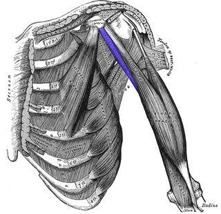
The biceps or biceps brachii is a large muscle that lies on the front of the upper arm between the shoulder and the elbow. Both heads of the muscle arise on the scapula and join to form a single muscle belly which is attached to the upper forearm. While the long head of the biceps crosses both the shoulder and elbow joints, its main function is at the elbow where it flexes and supinates the forearm. Both these movements are used when opening a bottle with a corkscrew: first biceps screws in the cork (supination), then it pulls the cork out (flexion).

The brachioradialis is a muscle of the forearm that flexes the forearm at the elbow. It is also capable of both pronation and supination, depending on the position of the forearm. It is attached to the distal styloid process of the radius by way of the brachioradialis tendon, and to the lateral supracondylar ridge of the humerus.

The radial nerve is a nerve in the human body that supplies the posterior portion of the upper limb. It innervates the medial and lateral heads of the triceps brachii muscle of the arm, as well as all 12 muscles in the posterior osteofascial compartment of the forearm and the associated joints and overlying skin.
The forearm is the region of the upper limb between the elbow and the wrist. The term forearm is used in anatomy to distinguish it from the arm, a word which is used to describe the entire appendage of the upper limb, but which in anatomy, technically, means only the region of the upper arm, whereas the lower "arm" is called the forearm. It is homologous to the region of the leg that lies between the knee and the ankle joints, the crus.

The ulnar nerve is a nerve that runs near the ulna, one of the two long bones in the forearm. The ulnar collateral ligament of elbow joint is in relation with the ulnar nerve. The nerve is the largest in the human body unprotected by muscle or bone, so injury is common. This nerve is directly connected to the little finger, and the adjacent half of the ring finger, innervating the palmar aspect of these fingers, including both front and back of the tips, perhaps as far back as the fingernail beds.

The upper limbs or upper extremities are the forelimbs of an upright-postured tetrapod vertebrate, extending from the scapulae and clavicles down to and including the digits, including all the musculatures and ligaments involved with the shoulder, elbow, wrist and knuckle joints. In humans, each upper limb is divided into the shoulder, arm, elbow, forearm, wrist and hand, and is primarily used for climbing, lifting and manipulating objects. In anatomy, just as arm refers to the upper arm, leg refers to the lower leg.

The triceps, or triceps brachii, is a large muscle on the back of the upper limb of many vertebrates. It consists of three parts: the medial, lateral, and long head. It is the muscle principally responsible for extension of the elbow joint.

The cubital fossa, antecubital fossa, chelidon, or inside of elbow is the area on the anterior side of the upper part between the arm and forearm of a human or other hominid animals. It lies anteriorly to the elbow (antecubital) when in standard anatomical position. The cubital fossa is a triangular area having three borders.

The anconeus muscle is a small muscle on the posterior aspect of the elbow joint.
The coracobrachialis muscle muscle in the upper medial part of the arm. It is located within the anterior compartment of the arm. It originates from the coracoid process of the scapula; it inserts onto the middle of the medial aspect of the body of the humerus. It is innervated by the musculocutaneous nerve. It acts to adduct and flex the arm.
The pronator teres is a muscle that, along with the pronator quadratus, serves to pronate the forearm. It has two origins, at the medial humeral supracondylar ridge and the medial side of the coronoid process of the ulna and inserts near the middle of the radius.

The inferior ulnar collateral artery is an artery in the arm. It arises about 5 cm. above the elbow from the brachial artery.
The superior ulnar collateral artery, of small size, arises from the brachial artery a little below the middle of the arm; it frequently springs from the upper part of the a. profunda brachii.
The deep artery of arm is a large artery of the arm which arises from the brachial artery. It descends in the arm before ending by anastomosing with the radial recurrent artery.
The brachial fascia is continuous with that covering the deltoideus and the pectoralis major muscle, by means of which it is attached, above, to the clavicle, acromion, and spine of the scapula; it forms a thin, loose, membranous sheath for the muscles of the arm, and sends septa between them; it is composed of fibers disposed in a circular or spiral direction, and connected together by vertical and oblique fibers.

The humeroulnar joint is part of the elbow-joint. It is composed of two bones, the humerus and ulna, and is the junction between the trochlear notch of ulna and the trochlea of humerus. It is classified as a simple hinge-joint, which allows for movements of flexion, extension and circumduction. Owing to the obliquity of the trochlea of the humerus, this movement does not take place in the antero-posterior plane of the body of the humerus.

The posterior compartment of the thigh is one of the fascial compartments that contains the knee flexors and hip extensors known as the hamstring muscles, as well as vascular and nervous elements, particularly the sciatic nerve.

The elbow is the region between the upper arm and the forearm that surrounds the elbow joint. The elbow includes prominent landmarks such as the olecranon, the cubital fossa, and the lateral and the medial epicondyles of the humerus. The elbow joint is a hinge joint between the arm and the forearm; more specifically between the humerus in the upper arm and the radius and ulna in the forearm which allows the forearm and hand to be moved towards and away from the body. The term elbow is specifically used for humans and other primates, and in other vertebrates it is not used. In those cases, forelimb plus joint is used.

