Brachial artery

Last updated

Brachial artery
Gray525.png
The brachial artery
Gray526.png
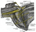
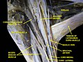
Right upper limb, anterior view, brachial artery and elbow.
Details
Source axillary artery
Branches Profunda brachii
Superior ulnar collateral artery
Inferior ulnar collateral artery
Radial artery
Ulnar artery
Vein brachial vein
Supplies biceps brachii muscle, triceps brachii muscle, coracobrachialis
Identifiers
Latin arteria brachialis
MeSH D001916
TA98 A12.2.09.018
TA2 4632
FMA 22689
Anatomical terminology

The brachial artery is the major blood vessel of the (upper) arm. It is the continuation of the axillary artery beyond the lower margin of teres major muscle. It continues down the ventral surface of the arm until it reaches the cubital fossa at the elbow. It then divides into the radial and ulnar arteries which run down the forearm. [1] [2] In some individuals, the bifurcation occurs much earlier and the ulnar and radial arteries extend through the upper arm. The pulse of the brachial artery is palpable on the anterior aspect of the elbow, medial to the tendon of the biceps, and, with the use of a stethoscope and sphygmomanometer (blood pressure cuff), often used to measure the blood pressure. [1]

Contents

The brachial artery is closely related to the median nerve; in proximal regions, the median nerve is immediately lateral to the brachial artery. Distally, the median nerve crosses the medial side of the brachial artery and lies anterior to the elbow joint. [1] [3]

Structure

The brachial artery gives rise to the following branches: [3]

It also gives rise to important anastomotic networks of the elbow and (as the axillary artery) the shoulder.

The biceps head is lateral to the brachial artery. The median nerve is medial to the brachial artery for most of its course.

Additional images

See also

Related Research Articles

<span class="mw-page-title-main">Arm</span> Proximal part of the free upper limb between the shoulder and the elbow

In human anatomy, the arm refers to the upper limb in common usage, although academically the term specifically means the upper arm between the glenohumeral joint and the elbow joint. The distal part of the upper limb between the elbow and the radiocarpal joint is known as the forearm or "lower" arm, and the extremity beyond the wrist is the hand.

<span class="mw-page-title-main">Humerus</span> Long bone of the upper arm

The humerus is a long bone in the arm that runs from the shoulder to the elbow. It connects the scapula and the two bones of the lower arm, the radius and ulna, and consists of three sections. The humeral upper extremity consists of a rounded head, a narrow neck, and two short processes. The body is cylindrical in its upper portion, and more prismatic below. The lower extremity consists of 2 epicondyles, 2 processes, and 3 fossae. As well as its true anatomical neck, the constriction below the greater and lesser tubercles of the humerus is referred to as its surgical neck due to its tendency to fracture, thus often becoming the focus of surgeons.

<span class="mw-page-title-main">Radial nerve</span> Nerve in the human body that supplies the posterior portion of the upper limb

The radial nerve is a nerve in the human body that supplies the posterior portion of the upper limb. It innervates the medial and lateral heads of the triceps brachii muscle of the arm, as well as all 12 muscles in the posterior osteofascial compartment of the forearm and the associated joints and overlying skin.

<span class="mw-page-title-main">Median nerve</span> Nerve of the upper limb

The median nerve is a nerve in humans and other animals in the upper limb. It is one of the five main nerves originating from the brachial plexus.

<span class="mw-page-title-main">Brachial plexus</span> Network of nerves

The brachial plexus is a network of nerves formed by the anterior rami of the lower four cervical nerves and first thoracic nerve. This plexus extends from the spinal cord, through the cervicoaxillary canal in the neck, over the first rib, and into the armpit, it supplies afferent and efferent nerve fibers to the chest, shoulder, arm, forearm, and hand.

The forearm is the region of the upper limb between the elbow and the wrist. The term forearm is used in anatomy to distinguish it from the arm, a word which is used to describe the entire appendage of the upper limb, but which in anatomy, technically, means only the region of the upper arm, whereas the lower "arm" is called the forearm. It is homologous to the region of the leg that lies between the knee and the ankle joints, the crus.

<span class="mw-page-title-main">Ulnar nerve</span> Nerve which runs near the ulna bone

In human anatomy, the ulnar nerve is a nerve that runs near the ulna bone. The ulnar collateral ligament of elbow joint is in relation with the ulnar nerve. The nerve is the largest in the human body unprotected by muscle or bone, so injury is common. This nerve is directly connected to the little finger, and the adjacent half of the ring finger, innervating the palmar aspect of these fingers, including both front and back of the tips, perhaps as far back as the fingernail beds.

<span class="mw-page-title-main">Upper limb</span> Consists of the arm, forearm, and hand

The upper limbs or upper extremities are the forelimbs of an upright-postured tetrapod vertebrate, extending from the scapulae and clavicles down to and including the digits, including all the musculatures and ligaments involved with the shoulder, elbow, wrist and knuckle joints. In humans, each upper limb is divided into the arm, forearm and hand, and is primarily used for climbing, lifting and manipulating objects.

<span class="mw-page-title-main">Radial artery</span> Large forearm artery

In human anatomy, the radial artery is the main artery of the lateral aspect of the forearm.

<span class="mw-page-title-main">Cubital fossa</span> The human elbow pit

The cubital fossa,chelidon, grivet or elbow pit, is the triangular area on the anterior side of the upper limb between the arm and forearm of a human or other hominid animals. It lies anteriorly to the elbow when in standard anatomical position.

<span class="mw-page-title-main">Basilic vein</span> Large blood vessel in the arm

The basilic vein is a large superficial vein of the upper limb that helps drain parts of the hand and forearm. It originates on the medial (ulnar) side of the dorsal venous network of the hand and travels up the base of the forearm, where its course is generally visible through the skin as it travels in the subcutaneous fat and fascia lying superficial to the muscles. The basilic vein terminates by uniting with the brachial veins to form the axillary vein.

<span class="mw-page-title-main">Ulnar artery</span> Artery of the forearm

The ulnar artery is the main blood vessel, with oxygenated blood, of the medial aspects of the forearm. It arises from the brachial artery and terminates in the superficial palmar arch, which joins with the superficial branch of the radial artery. It is palpable on the anterior and medial aspect of the wrist.

The pronator teres is a muscle that, along with the pronator quadratus, serves to pronate the forearm. It has two attachments, to the medial humeral supracondylar ridge and the ulnar tuberosity, and inserts near the middle of the radius.

<span class="mw-page-title-main">Medial cutaneous nerve of forearm</span>

The medial cutaneous nerve of the forearm is a sensory branch of the medial cord of the brachial plexus derived from the ventral rami of spinal nerves C8-T1. It provides sensory innervation to the skin of the medial forearm and skin overlying the olecranon. It descends through the (upper) arm within the brachial fascia alongside the basilic vein, then divides into an anterior branch and a posterior branch upon emerging from the brachial fascia; the two terminal branches travel as far distally as the wrist.

<span class="mw-page-title-main">Medial cutaneous nerve of arm</span>

The medial brachial cutaneous nerve is a sensory branch of the medial cord of the brachial plexus derived from spinal nerves C8-T1. It provides sensory innervation to the medial arm. It descends accompanied by the basilic vein.

<span class="mw-page-title-main">Bicipital aponeurosis</span>

The bicipital aponeurosis is a broad aponeurosis of the biceps brachii, which is located in the cubital fossa of the elbow. It separates superficial from deep structures in much of the fossa.

<span class="mw-page-title-main">Fascial compartments of arm</span> Anatomical compartments

The fascial compartments of arm refers to the specific anatomical term of the compartments within the upper segment of the upper limb of the body. The upper limb is divided into two segments, the arm and the forearm. Each of these segments is further divided into two compartments which are formed by deep fascia – tough connective tissue septa (walls). Each compartment encloses specific muscles and nerves.

<span class="mw-page-title-main">Outline of human anatomy</span> Overview of and topical guide to human anatomy

The following outline is provided as an overview of and topical guide to human anatomy:

<span class="mw-page-title-main">Elbow</span> Joint between the upper and lower parts of the arm

The elbow is the region between the upper arm and the forearm that surrounds the elbow joint. The elbow includes prominent landmarks such as the olecranon, the cubital fossa, and the lateral and the medial epicondyles of the humerus. The elbow joint is a hinge joint between the arm and the forearm; more specifically between the humerus in the upper arm and the radius and ulna in the forearm which allows the forearm and hand to be moved towards and away from the body. The term elbow is specifically used for humans and other primates, and in other vertebrates forelimb plus joint is used.

References

  1. 1 2 3 Gray's anatomy : the anatomical basis of clinical practice. Standring, Susan (Forty-first ed.). [Philadelphia]. 2016. ISBN   9780702052309. OCLC   920806541.{{cite book}}: CS1 maint: others (link)
  2. "Brachial artery". Kenhub. Retrieved September 28, 2019.
  3. 1 2 Moore, Keith L. (February 13, 2013). Clinically oriented anatomy. Dalley, Arthur F., II,, Agur, A. M. R. (Seventh ed.). Philadelphia. ISBN   978-1451119459. OCLC   813301028.