First dorsal metacarpal artery - arises just before the radial artery passes between the two heads of the first dorsal interosseous muscle and divides almost immediately into two branches which supply the adjacent sides of the thumb and index finger; the lateral side of the thumb receives a branch directly from the radial artery.
In the hand
Princeps pollicis artery - arises from the radial artery just as it turns medially to the deep part of the hand.
Radialis indicis - arises close to the princeps pollicis. The two arteries may arise from a common trunk, the first palmar metacarpal artery.
In less than 1% of the population, the radial artery takes a superficial course in the anatomical snuff box.[1] This arterial variation can be mistaken for the cephalic vein as accidental injection of this variant radial artery has been reported.[2] Identifying arterial pulsation in the anatomical snuff box is therefore recommended.
Clinical significance
The radial artery lies superficially in front of the distal end of the radius, between the tendons of the brachioradialis and flexor carpi radialis; it is here (the so-called gouttière du pouls) that clinician takes the radial pulse. (where it is commonly used to assess the heart rate and cardiac rhythm). Presence of radial pulse was thought to indicate a systolic blood pressure of at least 70 mmHg, as estimated from the 50% percentile, although this was found to generally be an overestimation of a patient's true blood pressure.[3] The radial artery can be less easily felt as it crosses the anatomical snuff box. The radial artery is used for coronary artery bypass grafting and is growing in popularity among cardiac surgeons.[4] Recently, it has been shown to have a superior peri-operative and post-operative course when compared to saphenous vein grafts.[5]
The radial artery is also used to evaluate the collateral circulation of blood through the hands; applying pressure through palpating the palmar arches results in paleness over the area being compressed; adequate collateral circulation can be ascertained by how quickly normal colour returns after the pressure is removed.[6]
The radial artery is a common site for the insertion of an arterial line, such as for blood pressure monitoring in an intensive care unit. It is also commonly used for cerebral angiograms for the treatment of cerebral pathologies, such as strokes,[7] cerebral aneurysms [8] It is selected because it is accessible, and because of the low incidence of complications such as thrombosis.[9]
Additional images
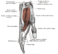
Surgical relations of the radial and ulnar arteries. Right upper limb. Anterior view. (After Gerrish.)
Cross-section through the middle of the right forearm.
The radial and ulnar arteries.
Front of right upper extremity, showing surface markings for bones, arteries, and nerves.
↑Fuhrman, Thomas M.; Pippin, William D.; Talmage, Lance A.; Reilley, Thomas E. (January 1, 1992). "Evaluation of collateral circulation of the hand". Journal of Clinical Monitoring. 8 (1): 28–32. doi:10.1007/BF01618084. ISSN1573-2614. PMID1538249. S2CID31956149.
↑Mouchtouris, Nikolaos; Al Saiegh, Fadi; Sweid, Ahmad; Amllay, Abdelaziz; Tjoumakaris, Stavropoula; Gooch, Reid; Rosenwasser, Robert; Jabbour, Pascal M. (November 2019). "Transradial Access for Newly Food and Drug Administration–Approved Devices for Endovascular Treatment of Cerebral Aneurysms: A Technical Note". World Neurosurgery. 131: 6–9. doi:10.1016/j.wneu.2019.07.149.
↑Bersten, Andrew D (2013). Oh's Intensive Care Manual. p.123. ISBN9780702047626.
This page is based on this Wikipedia article Text is available under the CC BY-SA 4.0 license; additional terms may apply. Images, videos and audio are available under their respective licenses.