The radial nerve is a nerve in the human body that supplies the posterior portion of the upper limb. It innervates the medial and lateral heads of the triceps brachii muscle of the arm, as well as all 12 muscles in the posterior osteofascial compartment of the forearm and the associated joints and overlying skin.
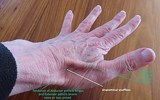
The anatomical snuff box or snuffbox or foveola radialis is a triangular deepening on the radial, dorsal aspect of the hand—at the level of the carpal bones, specifically, the scaphoid and trapezium bones forming the floor. The name originates from the use of this surface for placing and then sniffing powdered tobacco, or "snuff." It is sometimes referred to by its French name tabatière.

In human anatomy, the ulnar nerve is a nerve that runs near the ulna bone. The ulnar collateral ligament of elbow joint is in relation with the ulnar nerve. The nerve is the largest in the human body unprotected by muscle or bone, so injury is common. This nerve is directly connected to the little finger, and the adjacent half of the ring finger, innervating the palmar aspect of these fingers, including both front and back of the tips, perhaps as far back as the fingernail beds.

The scaphoid bone is one of the carpal bones of the wrist. It is situated between the hand and forearm on the thumb side of the wrist. It forms the radial border of the carpal tunnel. The scaphoid bone is the largest bone of the proximal row of wrist bones, its long axis being from above downward, lateralward, and forward. It is approximately the size and shape of a medium cashew nut.

The upper limbs or upper extremities are the forelimbs of an upright-postured tetrapod vertebrate, extending from the scapulae and clavicles down to and including the digits, including all the musculatures and ligaments involved with the shoulder, elbow, wrist and knuckle joints. In humans, each upper limb is divided into the arm, forearm and hand, and is primarily used for climbing, lifting and manipulating objects.

In human anatomy, the radial artery is the main artery of the lateral aspect of the forearm.
The ulnar artery is the main blood vessel, with oxygenated blood, of the medial aspects of the forearm. It arises from the brachial artery and terminates in the superficial palmar arch, which joins with the superficial branch of the radial artery. It is palpable on the anterior and medial aspect of the wrist.
In human anatomy, the extensor pollicis longus muscle (EPL) is a skeletal muscle located dorsally on the forearm. It is much larger than the extensor pollicis brevis, the origin of which it partly covers and acts to stretch the thumb together with this muscle.
In human anatomy, the extensor pollicis brevis (EPB) is a skeletal muscle on the dorsal side of the forearm. It lies on the medial side of, and is closely connected with, the abductor pollicis longus. The extensor pollicis brevis belongs to the deep group of the posterior fascial compartment of the forearm. It is a part of the lateral border of the anatomical snuffbox.

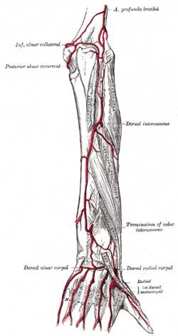
The posterior interosseous artery is an artery of the forearm. It is a branch of the common interosseous artery, which is a branch of the ulnar artery.
The anterior interosseous artery is an artery in the forearm. It is a branch of the common interosseous artery.
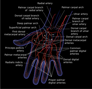
The superficial palmar arch is formed predominantly by the ulnar artery, with a contribution from the superficial palmar branch of the radial artery. However, in some individuals the contribution from the radial artery might be absent, and instead anastomoses with either the princeps pollicis artery, the radialis indicis artery, or the median artery, the former two of which are branches from the radial artery.
The deep palmar arch is an arterial network found in the palm. It is usually primarily formed from the terminal part of the radial artery. The ulnar artery also contributes through an anastomosis. This is in contrast to the superficial palmar arch, which is formed predominantly by the ulnar artery.
The princeps pollicis artery, or principal artery of the thumb, arises from the radial artery just as it turns medially towards the deep part of the hand; it descends between the first dorsal interosseous muscle and the oblique head of the adductor pollicis, along the medial side of the first metacarpal bone to the base of the proximal phalanx, where it lies beneath the tendon of the flexor pollicis longus muscle and divides into two branches.
The radialis indicis artery is a branch of the radial artery that provides blood to the index finger.
The dorsal carpal arch is an anatomical term for the combination (anastomosis) of dorsal carpal branch of the radial artery and the dorsal carpal branch of the ulnar artery near the back of the wrist.

Most of the dorsal metacarpal arteries arise from the dorsal carpal arch and run downward on the second, third, and fourth dorsal interossei of the hand and bifurcate into the dorsal digital arteries. Near their origin, they anastomose with the deep palmar arch by perforating arteries. They also anastomose with common palmar digital arteries, also via perforating arteries.
In anatomy, arterial tree is used to refer to all arteries and/or the branching pattern of the arteries. This article regards the human arterial tree. Starting from the aorta:
The dorsal carpal branch of the ulnar artery arises from the ulnar artery immediately above the pisiform bone, and winds backward beneath the tendon of the flexor carpi ulnaris; it passes across the dorsal surface of the carpus beneath the extensor tendons, to anastomose with a corresponding branch of the radial artery.

The extrinsic extensor muscles of the hand are located in the back of the forearm and have long tendons connecting them to bones in the hand, where they exert their action. Extrinsic denotes their location outside the hand. Extensor denotes their action which is to extend, or open flat, joints in the hand. They include the extensor carpi radialis longus (ECRL), extensor carpi radialis brevis (ECRB), extensor digitorum (ED), extensor digiti minimi (EDM), extensor carpi ulnaris (ECU), abductor pollicis longus (APL), extensor pollicis brevis (EPB), extensor pollicis longus (EPL), and extensor indicis (EI).