Axillary artery

Last updated

Axillary artery
Axillary limits.PNG
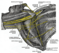
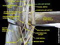
Axillary artery and its branches—anterior view of right upper limb and thorax. Upper and lower limits labeled.
Pectoralis minor.svg
The pectoralis minor muscle is used as a landmark for dividing the axillary artery into three parts.
Details
Source subclavian artery
Branches Superior thoracic
Thoracoacromial
Lateral thoracic
Subscapular
Anterior circumflex humeral
Posterior circumflex humeral
continues as brachial artery
Vein axillary vein
Supplies axilla
Identifiers
Latin arteria axillaris
MeSH D001366
TA98 A12.2.09.002
TA2 4616
FMA 22654
Anatomical terminology

In human anatomy, the axillary artery is a large blood vessel that conveys oxygenated blood to the lateral aspect of the thorax, the axilla (armpit) and the upper limb. Its origin is at the lateral margin of the first rib, before which it is called the subclavian artery.

Contents

After passing the lower margin of teres major it becomes the brachial artery.

Structure

The axillary artery is often referred to as having three parts, with these divisions based on its location relative to the Pectoralis minor muscle, which is superficial to the artery.

Relations

The axillary artery is accompanied by the axillary vein, [1] which lies medial to the artery, along its length.

In the axilla, the axillary artery is surrounded by the brachial plexus. [1] The second part of the axillary artery is the reference for the locational descriptions of the cords in the brachial plexus. For example, the posterior cord of the brachial plexus is so named because it lies posterior to the second part of the artery.

Branches

Branches of axillary artery Axillary branches.PNG
Branches of axillary artery

The axillary artery has several smaller branches. The branches can be remembered, in order, when traveling from the heart, with the mnemonics "Screw The Lawyers Save A Patient", "Summertime: The Lakers Schedule Another Parade", "Sixties Teens Love Sex And Pot", or "She Tastes Like Sweet Apple Pie." [2] The origin of these branches is highly variable (e.g. the posterior and anterior circumflex arteries often have a common trunk). An arterial branch is named for its course, not its origin.

Continues as the brachial artery past the inferior border of the teres major. [1]

Clinical significance

The axillary artery can be safely clamped without endangering the arm, but only in a location proximal to the origin of the subscapular artery (and distal to the thyrocervical trunk of the subclavian artery). The anastomotic network surrounding the scapula provides an alternate path for collateral circulation to the arm from arteries including the dorsal scapular artery and suprascapular artery.

The right axillary artery is often used as an arterial cannulation site in cardiac surgery, particularly for repair of aortic dissection and replacement of the ascending aorta and aortic arch.

Additional images

Related Research Articles

Arm Proximal part of the free upper limb between the shoulder and the elbow

In human anatomy, the arm is the part of the upper limb between the glenohumeral joint and the elbow joint. In common usage, the arm extends through the hand. The arm can be divided into the upper arm, which extends from the shoulder to the elbow, the forearm which extends from the elbow to the hand, and the hand. Anatomically the shoulder girdle with bones and corresponding muscles is by definition a part of the arm. The Latin term brachium may refer to either the arm as a whole or to the upper arm on its own.

Axillary nerve

The axillary nerve or the circumflex nerve is a nerve of the human body, that originates from the brachial plexus at the level of the axilla (armpit) and carries nerve fibers from C5 and C6. The axillary nerve travels through the quadrangular space with the posterior circumflex humeral artery and vein to innervate the deltoid and teres minor.

Subclavian artery Major arteries of the upper thorax, below the clavicle

In human anatomy, the subclavian arteries are paired major arteries of the upper thorax, below the clavicle. They receive blood from the aortic arch. The left subclavian artery supplies blood to the left arm and the right subclavian artery supplies blood to the right arm, with some branches supplying the head and thorax. On the left side of the body, the subclavian comes directly off the aortic arch, while on the right side it arises from the relatively short brachiocephalic artery when it bifurcates into the subclavian and the right common carotid artery.

Deltoid muscle Shoulder muscle

The deltoid muscle is the muscle forming the rounded contour of the human shoulder. It is also known as the 'common shoulder muscle', particularly in other animals such as the domestic cat. Anatomically, the deltoid muscle appears to be made up of three distinct sets of muscle fibers, namely the

  1. anterior or clavicular part
  2. posterior or scapular part
  3. intermediate or acromial part
Upper limb

The upper limbs or upper extremities are the forelimbs of an upright-postured tetrapod vertebrate, extending from the scapulae and clavicles down to and including the digits, including all the musculatures and ligaments involved with the shoulder, elbow, wrist and knuckle joints. In humans, each upper limb is divided into the arm, forearm and hand, and is primarily used for climbing, lifting and manipulating objects.

Axilla Area of the human body beneath the joint between arm and torso

The axilla is the area on the human body directly under the joint where the arm connects to the shoulder. It also contains many sweat glands.

Teres minor muscle

The teres minor is a narrow, elongated muscle of the rotator cuff. The muscle originates from the lateral border and adjacent posterior surface of the corresponding right or left scapula and inserts at both the greater tubercle of the humerus and the posterior surface of the joint capsule.

Axillary vein Large vein between the thorax and the heart

In human anatomy, the axillary vein is a large blood vessel that conveys blood from the lateral aspect of the thorax, axilla (armpit) and upper limb toward the heart. There is one axillary vein on each side of the body.

Lateral thoracic artery Large blood vessel

In human anatomy, the lateral thoracic artery is a blood vessel that supplies oxygenated blood to the lateral structures of the thorax and breast.

Shoulder joint Synovial ball and socket joint in the shoulder

The shoulder joint is structurally classified as a synovial ball-and-socket joint and functionally as a diarthrosis and multiaxial joint. It involves an articulation between the glenoid fossa of the scapula and the head of the humerus. Due to the very loose joint capsule that gives a limited interface of the humerus and scapula, it is the most mobile joint of the human body.

Subclavius muscle

The subclavius is a small triangular muscle, placed between the clavicle and the first rib. Along with the pectoralis major and pectoralis minor muscles, the subclavius muscle makes up the anterior axioappendicular muscles, also known as anterior wall of the axilla.

Medial pectoral nerve

The medial pectoral nerve arises from the medial cord of the brachial plexus, and through it from the eighth cervical and first thoracic roots (C8/T1).

Circumflex scapular artery Artery of the upper body

The circumflex scapular artery is a branch of the subscapular artery and part of the scapular anastomoses.

Transverse cervical artery Artery of the neck

The transverse cervical artery is an artery in the neck and a branch of the thyrocervical trunk, running at a higher level than the suprascapular artery.

Suprascapular artery Artery of the neck

The suprascapular artery is a branch of the thyrocervical trunk on the neck.

Posterior humeral circumflex artery

The posterior humeral circumflex artery arises from the third part of axillary artery at the lower border of the subscapularis, and runs posteriorly with the axillary nerve through the quadrangular space.

Scapular anastomosis Circulatory system around the shoulder blade

The scapular anastomosis is a system connecting certain subclavian artery and their corresponding axillary artery, forming a circulatory anastomosis around the scapula. It allows blood to flow past the joint in case of occlusion, damage, or pinching of the following scapular arteries:

Axillary space

The axillary spaces are anatomic spaces. through which axillary contents leave the axilla. They consist of the quadrangular space, triangular space, and triangular interval. It is bounded by teres major, teres minor, medial border of the humerus, and long head of triceps brachii.

Outline of human anatomy Overview of and topical guide to human anatomy

The following outline is provided as an overview of and topical guide to human anatomy:

References

  1. 1 2 3 4 5 6 7 8 9 Ryan, Stephanie (2011). "Chapter 7". Anatomy for diagnostic imaging (Third ed.). Elsevier Ltd. p. 277. ISBN   9780702029714.
  2. MedicalMnemonics.com: 1208 852 663