Posterior humeral circumflex artery

Last updated
Posterior humeral circumflex artery
Axillary branches.PNG
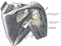
The axillary artery and its branches, including posterior humeral circumflex.
Gray524.png
The scapular and circumflex arteries. (Posterior hum. circumflex labeled at center right.)
Details
Source axillary artery
Identifiers
Latin arteria circumflexa humeri posterior
TA98 A12.2.09.017
TA2 4631
FMA 22684
Anatomical terminology

The posterior humeral circumflex artery (posterior circumflex artery or posterior circumflex humeral artery[ citation needed ]) arises from the third part of the axillary artery at the distal border of the subscapularis. [1]

Contents

Anatomy

Course and relations

It passes posteriorward with the axillary nerve through the quadrangular space. It winds laterally around the surgical neck of the humerus. [1]

Distribution

It is distributed to the shoulder joint, [1] teres major, teres minor, deltoid, [1] [2] and (long and lateral heads of) triceps brachii. [1]

Anastomoses

It forms anastomoses with the anterior humeral circumflex artery, (deltoid branch of) profunda brachii artery, (acromial branches of) suprascapular artery, (acromial branches of) and thoracoacromial artery. [1]

Additional images

See also

Related Research Articles

<span class="mw-page-title-main">Arm</span> Proximal part of the free upper limb between the shoulder and the elbow

In human anatomy, the arm refers to the upper limb in common usage, although academically the term specifically means the upper arm between the glenohumeral joint and the elbow joint. The distal part of the upper limb between the elbow and the radiocarpal joint is known as the forearm or "lower" arm, and the extremity beyond the wrist is the hand.

<span class="mw-page-title-main">Scapula</span> Bone that connects the humerus (upper arm bone) with the clavicle (collar bone)

The scapula, also known as the shoulder blade, is the bone that connects the humerus with the clavicle. Like their connected bones, the scapulae are paired, with each scapula on either side of the body being roughly a mirror image of the other. The name derives from the Classical Latin word for trowel or small shovel, which it was thought to resemble.

<span class="mw-page-title-main">Humerus</span> Long bone of the upper arm

The humerus is a long bone in the arm that runs from the shoulder to the elbow. It connects the scapula and the two bones of the lower arm, the radius and ulna, and consists of three sections. The humeral upper extremity consists of a rounded head, a narrow neck, and two short processes. The body is cylindrical in its upper portion, and more prismatic below. The lower extremity consists of 2 epicondyles, 2 processes, and 3 fossae. As well as its true anatomical neck, the constriction below the greater and lesser tubercles of the humerus is referred to as its surgical neck due to its tendency to fracture, thus often becoming the focus of surgeons.

<span class="mw-page-title-main">Brachial artery</span> Large blood vessel

The brachial artery is the major blood vessel of the (upper) arm. It is the continuation of the axillary artery beyond the lower margin of teres major muscle. It continues down the ventral surface of the arm until it reaches the cubital fossa at the elbow. It then divides into the radial and ulnar arteries which run down the forearm. In some individuals, the bifurcation occurs much earlier and the ulnar and radial arteries extend through the upper arm. The pulse of the brachial artery is palpable on the anterior aspect of the elbow, medial to the tendon of the biceps, and, with the use of a stethoscope and sphygmomanometer, often used to measure the blood pressure.

<span class="mw-page-title-main">Axillary nerve</span> Nerve of the human body near the armpit

The axillary nerve or the circumflex nerve is a nerve of the human body, that originates from the brachial plexus at the level of the axilla (armpit) and carries nerve fibers from C5 and C6. The axillary nerve travels through the quadrangular space with the posterior circumflex humeral artery and vein to innervate the deltoid and teres minor.

<span class="mw-page-title-main">Deltoid muscle</span> Shoulder muscle

The deltoid muscle is the muscle forming the rounded contour of the human shoulder. It is also known as the 'common shoulder muscle', particularly in other animals such as the domestic cat. Anatomically, the deltoid muscle appears to be made up of three distinct sets of muscle fibers, namely the

  1. anterior or clavicular part
  2. posterior or scapular part
  3. intermediate or acromial part
<span class="mw-page-title-main">Teres minor muscle</span> Muscle of the rotator cuff

The teres minor is a narrow, elongated muscle of the rotator cuff. The muscle originates from the lateral border and adjacent posterior surface of the corresponding right or left scapula and inserts at both the greater tubercle of the humerus and the posterior surface of the joint capsule.

<span class="mw-page-title-main">Axillary artery</span> Large blood vessel bringing oxygenated blood to the thorax

In human anatomy, the axillary artery is a large blood vessel that conveys oxygenated blood to the lateral aspect of the thorax, the axilla (armpit) and the upper limb. Its origin is at the lateral margin of the first rib, before which it is called the subclavian artery.

<span class="mw-page-title-main">Axillary vein</span> Large vein between the thorax and the heart

In human anatomy, the axillary vein is a large blood vessel that conveys blood from the lateral aspect of the thorax, axilla (armpit) and upper limb toward the heart. There is one axillary vein on each side of the body.

<span class="mw-page-title-main">Shoulder joint</span> Synovial ball and socket joint in the shoulder

The shoulder joint is structurally classified as a synovial ball-and-socket joint and functionally as a diarthrosis and multiaxial joint. It involves an articulation between the glenoid fossa of the scapula and the head of the humerus. Due to the very loose joint capsule that gives a limited interface of the humerus and scapula, it is the most mobile joint of the human body.

<span class="mw-page-title-main">Circumflex scapular artery</span> Artery of the upper body

The circumflex scapular artery is a branch of the subscapular artery and part of the scapular anastomoses.

<span class="mw-page-title-main">Suprascapular artery</span> Artery of the neck

The suprascapular artery is a branch of the thyrocervical trunk on the neck.

<span class="mw-page-title-main">Deep artery of arm</span> Deep arterial system of the arm

The deep artery of arm is a large artery of the arm which arises from the brachial artery. It descends in the arm before ending by anastomosing with the radial recurrent artery.

<span class="mw-page-title-main">Anterior humeral circumflex artery</span>

The anterior humeral circumflex artery is an artery in the arm. It is one of two circumflexing arteries that branch from the axillary artery, the other being the posterior humeral circumflex artery. The anterior humeral circumflex artery is considerably smaller than the posterior and arises nearly opposite to it, from the lateral side of the axillary artery.

In anatomy, arterial tree is used to refer to all arteries and/or the branching pattern of the arteries. This article regards the human arterial tree. Starting from the aorta:

<span class="mw-page-title-main">Surgical neck of the humerus</span> Part of one of the bones of the arm

The surgical neck of the humerus is a bony constriction at the proximal end of shaft of humerus. It is situated distal to the greater tubercle and lesser tubercle, and proximal to the deltoid tuberosity.

<span class="mw-page-title-main">Quadrangular space</span>

The quadrangular space, also known as the quadrilateral space (of Velpeau) and the foramen humerotricipitale, is one of the three spaces in the axillary space. The other two spaces are: triangular space and triangular interval.

<span class="mw-page-title-main">Triangular interval</span> Intermuscular space of the upper body

The triangular interval is a space found in the axilla. It is one of the three intermuscular spaces found in the axillary space. The other two spaces are: quadrangular space and triangular space.

<span class="mw-page-title-main">Axillary space</span>

The axillary spaces are anatomic spaces. through which axillary contents leave the axilla. They consist of the quadrangular space, triangular space, and triangular interval. It is bounded by teres major, teres minor, medial border of the humerus, and long head of triceps brachii.

<span class="mw-page-title-main">Outline of human anatomy</span> Overview of and topical guide to human anatomy

The following outline is provided as an overview of and topical guide to human anatomy:

References

  1. 1 2 3 4 5 6 Standring, Susan (2020). Gray's Anatomy: The Anatomical Basis of Clinical Practice (42nd ed.). New York. p. 921. ISBN   978-0-7020-7707-4. OCLC   1201341621.{{cite book}}: CS1 maint: location missing publisher (link)
  2. Clinically Oriented Anatomy 7th ed. 2014. p. 718.

PD-icon.svgThis article incorporates text in the public domain from page 589 of the 20th edition of Gray's Anatomy (1918)