The anterior branch (upper branch) winds around the surgical neck of the humerus,[5] beneath the deltoid muscle, with the posterior humeral circumflex vessels. It continues as far as the anterior border of the deltoid to provide motor innervation. The anterior branch also gives off a few small cutaneous branches, which pierce the muscle and supply in the overlaying skin.
The posterior branch (lower branch) supplies the teres minor and the posterior part of the deltoid.[2] The posterior branch pierces the deep fascia and continues as the superior (or upper) lateral cutaneous nerve of arm, which sweeps around the posterior border of the deltoid and supplies the skin over the lower two-thirds of the posterior part of this muscle, as well as that covering the long head of the triceps brachii.
The motor branch of the long head of the triceps brachii arises, on average, a distance of 6mm (range 2–12mm) from the terminal division of the posterior cord termination.[6]
The trunk of the axillary nerve gives off an articular filament which enters the shoulder joint below the subscapularis.
Variation
Traditionally, the axillary nerve is thought to only supply the deltoid and teres minor. However, several studies on cadavers pointed out that the long head of triceps brachii is innervated by a branch of the axillary nerve.[7][6][8]
The axillary nerve also carries sensory information from the shoulder joint. It also innervates the skin, covering the inferior region of the deltoid muscle, known as the regimental badge area.[9] This is innervated by the superior lateral cutaneous nerve branch of the axillary nerve.
The posterior cord of the brachial plexus splits inferiorly to the glenohumeral joint giving rise to the axillary nerve which wraps around the surgical neck of the humerus, and the radial nerve which wraps around the humerus anteriorly and descends along its lateral border.
Clinical significance
The axillary nerve may be injured in anterior-inferior dislocations of the shoulder joint, compression of the axilla with a crutch or fracture of the surgical neck of the humerus. An example of injury to the axillary nerve includes axillary nerve palsy. Injury to the nerve results in:
Paralysis of the teres minor muscle and deltoid muscle, resulting in loss of abduction of arm (from 15-90 degrees), weak flexion, extension, and rotation of shoulder. Paralysis of deltoid and teres minor muscles results in flat shoulder deformity.
Loss of sensation in the skin over the regimental badge area.[9]
Direct trauma to the nerve can also lead to paralysis and loss of sensation.[10]
Additional images
Brachial plexus with courses of spinal nerves shown
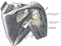
Suprascapular and axillary nerves of right side, seen from behind.
Cutaneous nerves of right upper extremity.
Diagram of segmental distribution of the cutaneous nerves of the right upper extremity.
1 2 de Sèze MP, Rezzouk J, de Sèze M, Uzel M, Lavignolle B, Midy D, Durandeau A (2004). "Does the motor branch of the long head of the triceps brachii arise from the radial nerve?". Surg Radiol Anat. 26 (6): 459–461. doi:10.1007/s00276-004-0253-z. PMID15365769. S2CID10052988.
↑ Rezzouk, J; Durandeau, A; Vital, JM; Fabre, T (October 2002). "Long head of the triceps brachii in axillary nerve injury: anatomy and clinical aspects". Revue de Chirurgie Orthopédique et Réparatrice de l'Appareil Moteur. 88 (6): 561–564. PMID12447125.
↑ Komala, Nanjundaiah; Shashanka, MallasandraJayadevaiah; Sheshgiri, Chowdapurkar (16 April 2012). "Long head of triceps supplied by axillary nerve". International Journal of Anatomical Variations. 5: 35–37. Retrieved 26 January 2018.
This page is based on this Wikipedia article Text is available under the CC BY-SA 4.0 license; additional terms may apply. Images, videos and audio are available under their respective licenses.