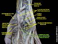
The radial nerve is a nerve in the human body that supplies the posterior portion of the upper limb. It innervates the medial and lateral heads of the triceps brachii muscle of the arm, as well as all 12 muscles in the posterior osteofascial compartment of the forearm and the associated joints and overlying skin.

The median nerve is a nerve in humans and other animals in the upper limb. It is one of the five main nerves originating from the brachial plexus.

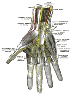
The ulnar nerve is a nerve that runs near the ulna, one of the two long bones in the forearm. The ulnar collateral ligament of elbow joint is in relation with the ulnar nerve. The nerve is the largest in the human body unprotected by muscle or bone, so injury is common. This nerve is directly connected to the little finger, and the adjacent half of the ring finger, innervating the palmar aspect of these fingers, including both front and back of the tips, perhaps as far back as the fingernail beds.

The upper limbs or upper extremities are the forelimbs of an upright-postured tetrapod vertebrate, extending from the scapulae and clavicles down to and including the digits, including all the musculatures and ligaments involved with the shoulder, elbow, wrist and knuckle joints. In humans, each upper limb is divided into the shoulder, arm, elbow, forearm, wrist and hand, and is primarily used for climbing, lifting and manipulating objects. In anatomy, just as arm refers to the upper arm, leg refers to the lower leg.

The tibial nerve is a branch of the sciatic nerve. The tibial nerve passes through the popliteal fossa to pass below the arch of soleus.

The superficial fibular nerve is a mixed nerve that provides motor innervation to the fibularis longus and fibularis brevis muscles, and sensory innervation to skin over the antero-lateral aspect of the leg along with the greater part of the dorsum of the foot.
The deep fibular nerve begins at the bifurcation of the common fibular nerve between the fibula and upper part of the fibularis longus, passes infero-medially, deep to the extensor digitorum longus, to the anterior surface of the interosseous membrane, and comes into relation with the anterior tibial artery above the middle of the leg; it then descends with the artery to the front of the ankle-joint, where it divides into a lateral and a medial terminal branch.

The lateral cutaneous nerve of forearm is a sensory nerve representing the continuation of the musculocutaneous nerve beyond the lateral edge of the tendon of the biceps brachii muscle. The lateral cutaneous nerve provides sensory innervation to the skin of the lateral forearm. It pierces the deep fascia of forearm to enter the subcutaneous compartment before splitting into a volar branch and a dorsal branch.
The medial cutaneous nerve of the forearm is a sensory branch of the medial cord of the brachial plexus derived from the ventral rami of spinal nerves C8-T1. It provides sensory innervation to the skin of the medial forearm and skin overlying the olecranon. It descends through the (upper) arm within the brachial fascia alongside the basilic vein, then divides into an anterior branch and a posterior branch upon emerging from the brachial fascia; the two terminal branches travel as far distally as the wrist.
The superficial branch of the radial nerve passes along the front of the radial side of the forearm to the commencement of its lower third. It is a sensory nerve.

The medial plantar nerve is the larger of the two terminal divisions of the tibial nerve, which accompanies the medial plantar artery.
The dorsal branch of ulnar nerve arises about 5 cm. proximal to the wrist; it passes backward beneath the Flexor carpi ulnaris, perforates the deep fascia, and, running along the ulnar side of the back of the wrist and hand, divides into two dorsal digital branches; one supplies the ulnar side of the little finger; the other, the adjacent sides of the little and ring fingers.

In the palm of the hand the median nerve is covered by the skin and the palmar aponeurosis, and rests on the tendons of the flexor muscles. Immediately after emerging from under the transverse carpal ligament the median nerve becomes enlarged and flattened and splits into a smaller, lateral, and a larger, medial portion.
The palmar branch of the median nerve is a branch of the median nerve which arises at the distal part of the forearm.
Cutaneous innervation of the upper limbs is the nerve supply to areas of the skin of the upper limbs which are supplied by specific cutaneous nerves.
The superficial branch of the lateral plantar nerve splits into a proper and a common plantar digital nerve:
The common plantar digital nerves of medial plantar nerve are nerves of the foot. The three common digital nerves pass between the divisions of the plantar aponeurosis, and each splits into two proper digital nerves:
The proper palmar digital nerves of the ulnar nerve are nerves of the hand.
The common palmar digital nerves of the ulnar nerve are nerves of the hand. The nerve branches off the superficial branch of the ulnar nerve and runs toward the cleft between the ring and little fingers.