Radial nerve

Last updated
Radial nerve
Gray818.png
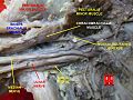
The suprascapular, axillary, and radial nerves.
Details
From posterior cord
To posterior interosseous nerve
Innervates posterior compartment of the arm, posterior compartment of the forearm
Identifiers
Latin nervus radialis
MeSH D011826
TA98 A14.2.03.049
TA2 6431
FMA 37069
Anatomical terms of neuroanatomy

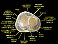
The radial nerve is a nerve in the human body that supplies the posterior portion of the upper limb. It innervates the medial and lateral heads of the triceps brachii muscle of the arm, as well as all 12 muscles in the posterior osteofascial compartment of the forearm and the associated joints and overlying skin.

Contents

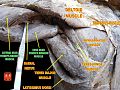
It originates from the brachial plexus, carrying fibers from the posterior roots of spinal nerves C5, C6, C7, C8 and T1. [1]

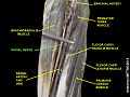
The radial nerve and its branches provide motor innervation to the dorsal arm muscles (the triceps brachii and the anconeus) and the extrinsic extensors of the wrists and hands; it also provides cutaneous sensory innervation to most of the back of the hand, except for the back of the little finger and adjacent half of the ring finger (which are innervated by the ulnar nerve).

The radial nerve divides into a deep branch, which becomes the posterior interosseous nerve, and a superficial branch, which goes on to innervate the dorsum (back) of the hand.

This nerve was historically referred to as the musculospiral nerve. [2]

Structure

Radial nerve of the right axilla, posterior view Radial nerve.jpg
Radial nerve of the right axilla, posterior view

The radial nerve originates as a terminal branch of the posterior cord of the brachial plexus. [1] It goes through the arm, first in the posterior compartment of the arm, and later in the anterior compartment of the arm, and continues in the posterior compartment of the forearm.

Arm

The radial nerve originates from the posterior cord of the brachial plexus with root values of C5 to C8 and T1. From the brachial plexus, it travels behind the third part of the axillary artery (part of the axillary artery distal to the pectoralis minor). In the arm, it runs behind the brachial artery and then enters the lower triangular space to reach the radial sulcus on the back of the humerus. [1] It travels downwards together with profunda brachii artery, between the lateral and medial heads of triceps brachii until it reaches the lateral side the arm at 5 cm below the deltoid tuberosity where it pierces the lateral intermuscular septum to reach the anterior compartment of the arm. Then, it descends down to cross the lateral epicondyle of the humerus where the nerve terminates by branching itself into superficial and deep branch which continues into cubital fossa and then into the forearm. [3]

Radial nerve gives out muscular branches to supply the long head, medial head, and lateral head of triceps brachii muscles before and during its course in the radial sulcus. After it emerges out from the radial sulcus, it supplies the brachialis, brachioradialis and extensor carpi radialis longus. [3]

Above the radial sulcus, the radial nerve gives off posterior cutaneous nerve of the arm which supplies the skin at the back of the arm. In the radial sulcus, it gives off lower lateral cutaneous nerve of the arm and posterior cutaneous nerve of the forearm. The radial nerve also gives articular branches to supply the elbow joint. [3]

Forearm and hand

In the forearm, it is divided into a superficial branch (primarily sensory) and a deep branch (primarily motor).

Variation

It is commonly believed that the radial nerve provides motor innervation to the long head of the triceps. However, a study conducted in 2004 found that axillary nerve innervated the long head of the triceps in twenty cadavers without any supply from the radial nerve. [6]

Function

The following are branches of the radial nerve (including the superficial branch of the radial nerve and the deep branch of the radial nerve/posterior interosseous nerve).

Cutaneous

Cutaneous innervation of the right upper extremity. Areas innervated by the radial nerve are colored in pink. Gray812and814.PNG
Cutaneous innervation of the right upper extremity. Areas innervated by the radial nerve are colored in pink.

Cutaneous innervation by the radial nerve is provided by the following nerve branches:

The superficial branch of the radial nerve provides sensory innervation to much of the back of the hand, including the web of skin between the thumb and index finger.

Motor

Muscles of the posterior forearm. All the labelled muscles (that is, all the visible muscles except the ones on the dorsal hand and one at top left) are innervated by the radial nerve, and represent all muscles innervated by the radial nerve except for the supinator. Gray418.png
Muscles of the posterior forearm. All the labelled muscles (that is, all the visible muscles except the ones on the dorsal hand and one at top left) are innervated by the radial nerve, and represent all muscles innervated by the radial nerve except for the supinator.

Muscular branches of the radial nerve:

Deep branch of the radial nerve:

Posterior interosseous nerve (a continuation of the deep branch after the supinator):

The radial nerve (and its deep branch) provides motor innervation to the muscles in the posterior compartment of the arm and forearm, which are mostly extensors.

Clinical significance

Injury

Injury to the radial nerve at different levels causes different syndromes with varying motor and sensory deficits.

At the axilla

At mid-arm

Just below the elbow

Within the distal forearm:

History

Additional images

See also

Related Research Articles

<span class="mw-page-title-main">Arm</span> Proximal part of the free upper limb between the shoulder and the elbow

In human anatomy, the arm refers to the upper limb in common usage, although academically the term specifically means the upper arm between the glenohumeral joint and the elbow joint. The distal part of the upper limb between the elbow and the radiocarpal joint is known as the forearm or "lower" arm, and the extremity beyond the wrist is the hand.

<span class="mw-page-title-main">Brachioradialis</span> Muscle of the upper limb

The brachioradialis is a muscle of the forearm that flexes the forearm at the elbow. It is also capable of both pronation and supination, depending on the position of the forearm. It is attached to the distal styloid process of the radius by way of the brachioradialis tendon, and to the lateral supracondylar ridge of the humerus.

<span class="mw-page-title-main">Median nerve</span> Nerve of the upper limb

The median nerve is a nerve in humans and other animals in the upper limb. It is one of the five main nerves originating from the brachial plexus.

The forearm is the region of the upper limb between the elbow and the wrist. The term forearm is used in anatomy to distinguish it from the arm, a word which is used to describe the entire appendage of the upper limb, but which in anatomy, technically, means only the region of the upper arm, whereas the lower "arm" is called the forearm. It is homologous to the region of the leg that lies between the knee and the ankle joints, the crus.

<span class="mw-page-title-main">Axillary nerve</span> Nerve of the human body near the armpit

The axillary nerve or the circumflex nerve is a nerve of the human body, that originates from the brachial plexus at the level of the axilla (armpit) and carries nerve fibers from C5 and C6. The axillary nerve travels through the quadrangular space with the posterior circumflex humeral artery and vein to innervate the deltoid and teres minor.

<span class="mw-page-title-main">Ulnar nerve</span> Nerve which runs near the ulna bone

In human anatomy, the ulnar nerve is a nerve that runs near the ulna bone. The ulnar collateral ligament of elbow joint is in relation with the ulnar nerve. The nerve is the largest in the human body unprotected by muscle or bone, so injury is common. This nerve is directly connected to the little finger, and the adjacent half of the ring finger, innervating the palmar aspect of these fingers, including both front and back of the tips, perhaps as far back as the fingernail beds.

<span class="mw-page-title-main">Wrist drop</span> Medical condition

Wrist drop is a medical condition in which the wrist and the fingers cannot extend at the metacarpophalangeal joints. The wrist remains partially flexed due to an opposing action of flexor muscles of the forearm. As a result, the extensor muscles in the posterior compartment remain paralyzed.

<span class="mw-page-title-main">Upper limb</span> Consists of the arm, forearm, and hand

The upper limbs or upper extremities are the forelimbs of an upright-postured tetrapod vertebrate, extending from the scapulae and clavicles down to and including the digits, including all the musculatures and ligaments involved with the shoulder, elbow, wrist and knuckle joints. In humans, each upper limb is divided into the arm, forearm and hand, and is primarily used for climbing, lifting and manipulating objects.

<span class="mw-page-title-main">Cubital fossa</span> The human elbow pit

The cubital fossa, chelidon or inside of elbow is the area on the anterior side of the upper part between the arm and forearm of a human or other hormid animals. It lies anteriorly to the elbow when in standard anatomical position.

In human anatomy, the extensor pollicis longus muscle (EPL) is a skeletal muscle located dorsally on the forearm. It is much larger than the extensor pollicis brevis, the origin of which it partly covers and acts to stretch the thumb together with this muscle.

<span class="mw-page-title-main">Supinator muscle</span> Muscle of the forearm in humans

In human anatomy, the supinator is a broad muscle in the posterior compartment of the forearm, curved around the upper third of the radius. Its function is to supinate the forearm.

<span class="mw-page-title-main">Posterior interosseous nerve</span> Nerve of the forearm

The posterior interosseous nerve is a nerve in the forearm. It is the continuation of the deep branch of the radial nerve, after this has crossed the supinator muscle. It is considerably diminished in size compared to the deep branch of the radial nerve. The nerve fibers originate from cervical segments C7 and C8 in the spinal column.

<span class="mw-page-title-main">Lateral cutaneous nerve of forearm</span>

The lateral cutaneous nerve of forearm is a sensory nerve representing the continuation of the musculocutaneous nerve beyond the lateral edge of the tendon of the biceps brachii muscle. The lateral cutaneous nerve provides sensory innervation to the skin of the lateral forearm. It pierces the deep fascia of forearm to enter the subcutaneous compartment before splitting into a volar branch and a dorsal branch.

<span class="mw-page-title-main">Deep artery of arm</span> Deep arterial system of the arm

The deep artery of arm is a large artery of the arm which arises from the brachial artery. It descends in the arm before ending by anastomosing with the radial recurrent artery.

<span class="mw-page-title-main">Deep branch of radial nerve</span>

The radial nerve divides into a superficial (sensory) and deep (motor) branch at the cubital fossa. The deep branch of the radial nerve winds to the back of the forearm around the lateral side of the radius between the two planes of fibers of the supinator, and is prolonged downward between the superficial and deep layers of muscles, to the middle of the forearm. The deep branch provides motor function to the muscles in the posterior compartment of the forearm, which is mostly the extensor muscles of the hand.

<span class="mw-page-title-main">Muscular branches of the radial nerve</span> Nerve system

The muscular branches of the radial nerve supply the Triceps brachii, Anconæus, Brachioradialis, and Extensor carpi radialis longus, and are grouped as medial, posterior, and lateral.

<span class="mw-page-title-main">Fascial compartments of arm</span> Anatomical compartments

The fascial compartments of arm refers to the specific anatomical term of the compartments within the upper segment of the upper limb of the body. The upper limb is divided into two segments, the arm and the forearm. Each of these segments is further divided into two compartments which are formed by deep fascia – tough connective tissue septa (walls). Each compartment encloses specific muscles and nerves.

The posterior compartment of the forearm contains twelve muscles which primarily extend the wrist and digits. It is separated from the anterior compartment by the interosseous membrane between the radius and ulna.

<span class="mw-page-title-main">Outline of human anatomy</span> Overview of and topical guide to human anatomy

The following outline is provided as an overview of and topical guide to human anatomy:

<span class="mw-page-title-main">Cervical spinal nerve 8</span> Spinal nerve of the cervical segment

The cervical spinal nerve 8 (C8) is a spinal nerve of the cervical segment.

References

  1. 1 2 3 4 Scott, Kevin R.; Ahmed, Aiesha; Scott, Linda; Kothari, Milind J. (2013-01-01), Barnes, Michael P.; Good, David C. (eds.), "Chapter 42 - Rehabilitation of brachial plexus and peripheral nerve disorders", Handbook of Clinical Neurology, Neurological Rehabilitation, 110, Elsevier: 499–514, doi:10.1016/b978-0-444-52901-5.00042-3, ISBN   9780444529015, PMID   23312667 , retrieved 2020-10-25
  2. Colborn, Gene L.; Goodrich, J. Alan; Levine, Monroe I.; Bhatti, Nazir A. (1993). "The variable anatomy of the nerve to the extensor carpi radialis brevis". Clinical Anatomy. 6 (1): 48–53. doi:10.1002/ca.980060108. ISSN   0897-3806. S2CID   72093080.
  3. 1 2 3 4 5 Krishna, Garg (2010). "8 - Arm". BD Chaurasia's Human Anatomy (Regional and Applied Dissection and Clinical) Volume 1 - Upper limb and thorax (5th ed.). India: CBS Publishers and Distributors Pvt Ltd. pp. 95, 111, 122, 128. ISBN   978-81-239-1863-1.
  4. "Acland's video Atlas of Human Anatomy 1.3.1.8 - Radial nerve in the forearm and hand". Wolters Kluwer. Archived from the original on 29 November 2017. Retrieved 26 January 2018. The deep branch of the radial nerve, also known as the posterior interosseous nerve, is a motor nerve.
  5. Nurul Huda, Mohd Nor; San, Aye Aye; Fauziah, Othman (2014). "An Anomalous Pattern of Superficial Branch of Radial Nerve: A Cadaveric Case Report". International Journal of Morphology. 32 (1): 29–31. doi: 10.4067/S0717-95022014000100005 . ISSN   0717-9502. The deep branch of radial nerve and also known as posterior interosseous nerve is the nerve of extensor compartment of the forearm ...
  6. de Sèze MP, Rezzouk J, de Sèze M, Uzel M, Lavignolle B, Midy D, Durandeau A (2004). "Does the motor branch of the long head of the triceps brachii arise from the radial nerve? An anatomic and electromyographic study". Surg Radiol Anat. 26 (6): 459–61. doi:10.1007/s00276-004-0253-z. PMID   15365769. S2CID   10052988.
  7. Ryan, Monique M.; Jones, H. Royden (2015-01-01), Darras, Basil T.; Jones, H. Royden; Ryan, Monique M.; De Vivo, Darryl C. (eds.), "Chapter 14 – Mononeuropathies", Neuromuscular Disorders of Infancy, Childhood, and Adolescence (Second Edition), San Diego: Academic Press, pp. 243–273, doi:10.1016/b978-0-12-417044-5.00014-7, ISBN   978-0-12-417044-5 , retrieved 2020-10-25
  8. 1 2 3 Prakash, M. V. S. Satya; Udupi Bidkar, Prasanna (2016-01-01), Prabhakar, Hemanshu (ed.), "Chapter 37 - Peripheral Nerve Injuries", Complications in Neuroanesthesia, San Diego: Academic Press, pp. 359–368, doi:10.1016/b978-0-12-804075-1.00037-7, ISBN   978-0-12-804075-1 , retrieved 2020-10-25
  9. 1 2 Kretschmer, Thomas; Heinen, Christian (2015-01-01), Tubbs, R. Shane; Rizk, Elias; Shoja, Mohammadali M.; Loukas, Marios (eds.), "Chapter 36 - Iatrogenic Injuries of the Nerves", Nerves and Nerve Injuries, San Diego: Academic Press, pp. 557–585, doi:10.1016/b978-0-12-802653-3.00085-3, ISBN   978-0-12-802653-3 , retrieved 2020-10-25
  10. Tor Wo Chiu (2011). Stone's Plastic Surgery Facts and Figures. Cambridge University Press. ISBN   9781139499781.