Radial tunnel syndrome

Last updated
Radial tunnel syndrome
Gray420.png
The supinator. (Deep branch of radial nerve labeled at upper right and lower right. Dorsal interosseous is this nerve after passing the supinator, at lower right.)
Specialty Neurology   OOjs UI icon edit-ltr-progressive.svg

Radial tunnel syndrome (RTS) is caused by increased pressure on the radial nerve as it travels from the upper arm (the brachial plexus) to the hand and wrist.

Contents

Symptoms and signs

Radial tunnel syndrome causes posterolateral elbow pain that is similar to tennis elbow and may sometimes occur in conjunction with that condition. Patients may typically have weakness of extension at the wrist and third digit. The pain is often reproduced upon resisted supination of the forearm, and pain at the radial tunnel on resisted hyperextension of the wrist.

Cause

The theory is that the radial nerve becomes irritated and/or inflamed from friction caused by compression by muscles in the forearm. [1]

Some speculate that radial tunnel syndrome is a type of repetitive strain injury (RSI), but there is no detectable pathophysiology and even the existence of this disorder is questioned. [1]

The term "radial tunnel syndrome" is used for compression of the posterior interosseous nerve, a division of the radial nerve, at the lateral intermuscular septum of arm, while "supinator syndrome" is used for compression at the arcade of Frohse. [2]

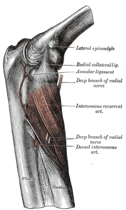
The "radial tunnel" is the region from the humeroradial joint past the proximal origin of the supinator muscle. Some scientists believe the radial tunnel extends as far as the distal border of the supinator. The radial nerve is commonly compressed within a 5 cm region near the elbow, but it can be compressed anywhere along the forearm if the syndrome is caused by injury (e.g. a fracture that puts pressure on the radial nerve). [3] The radial nerve provides sensation to the skin of posterior arm, posterior and lateral forearm and wrist, and the joints of the elbow, wrist and hand. The nerve also provides sensory branches that travel to the periosteum of the lateral epicondyle, the anterior radiohumeral joint, and the annular ligament. It provides motor function through innervation to most extensor muscles of the posterior arm and forearm. Therefore, it is extremely important in upper body extremity movement and can cause significant pain to patients presenting with radial tunnel syndrome. [4] Unlike carpal tunnel syndrome, radial tunnel syndrome does not present tingling or numbness, since the posterior interosseous nerve mainly affects motor function. [5]

This problem is often caused by: bone tumors, injury (specifically fractures of the forearm), noncancerous fatty tumors (lipomas), and inflammation of surrounding tissue. [5]

Diagnosis

The diagnosis is based on symptoms and signs alone and objective testing is expected to be normal. [1] This syndrome may be clinically tested by flexing the patients long finger while the patient extends the wrist and fingers. Pain is a positive finding.

The chief complaint of this disease is usually pain in the dorsal aspect of the upper forearm, and any weakness described is secondary to the pain. Tenderness to palpation occurs over the area of the radial neck. Also, the disease can be diagnosed by a positive "middle finger test", where resisted middle finger extension produces pain. Radiographic evaluation of the elbow should be performed to rule out other diagnoses. [3]

Treatment

Non-surgical treatment of radial tunnel syndrome includes rest, NSAID, therapy with modalities, work modification, ergonomic modification, injection if associated with lateral epicondylitis. [6]

Patients whose conditions are more adapted to surgical intervention are those who do not respond to prolonged conservative treatment. The patient must have pain with resisted supination, positive middle finger test, positive electrodiagnostic findings, and pain relief after anesthetic injection into the radial tunnel. Based on 2002 data, surgical decompression leads to 60–70% good or excellent results. [3]

See also

Related Research Articles

<span class="mw-page-title-main">Ulna</span> Medial bone from forearm

The ulna is a long bone found in the forearm that stretches from the elbow to the smallest finger, and when in anatomical position, is found on the medial side of the forearm. That is, the ulna is on the same side of the forearm as the little finger. It runs parallel to the radius, the other long bone in the forearm. The ulna is usually slightly longer than the radius, but the radius is thicker. Therefore, the radius is considered to be the larger of the two.

<span class="mw-page-title-main">Radial nerve</span> Nerve in the human body that supplies the posterior portion of the upper limb

The radial nerve is a nerve in the human body that supplies the posterior portion of the upper limb. It innervates the medial and lateral heads of the triceps brachii muscle of the arm, as well as all 12 muscles in the posterior osteofascial compartment of the forearm and the associated joints and overlying skin.

<span class="mw-page-title-main">Median nerve</span> Nerve of the upper limb

The median nerve is a nerve in humans and other animals in the upper limb. It is one of the five main nerves originating from the brachial plexus.

The flexor digitorum profundus is a muscle in the forearm of humans that flexes the fingers. It is considered an extrinsic hand muscle because it acts on the hand while its muscle belly is located in the forearm.

The forearm is the region of the upper limb between the elbow and the wrist. The term forearm is used in anatomy to distinguish it from the arm, a word which is used to describe the entire appendage of the upper limb, but which in anatomy, technically, means only the region of the upper arm, whereas the lower "arm" is called the forearm. It is homologous to the region of the leg that lies between the knee and the ankle joints, the crus.

<span class="mw-page-title-main">Ulnar nerve</span> Nerve which runs near the ulna bone

In human anatomy, the ulnar nerve is a nerve that runs near the ulna bone. The ulnar collateral ligament of elbow joint is in relation with the ulnar nerve. The nerve is the largest in the human body unprotected by muscle or bone, so injury is common. This nerve is directly connected to the little finger, and the adjacent half of the ring finger, innervating the palmar aspect of these fingers, including both front and back of the tips, perhaps as far back as the fingernail beds.

<span class="mw-page-title-main">Wrist drop</span> Medical condition

Wrist drop is a medical condition in which the wrist and the fingers cannot extend at the metacarpophalangeal joints. The wrist remains partially flexed due to an opposing action of flexor muscles of the forearm. As a result, the extensor muscles in the posterior compartment remain paralyzed.

<span class="mw-page-title-main">Radius (bone)</span> One of the two long bones of the forearm

The radius or radial bone is one of the two large bones of the forearm, the other being the ulna. It extends from the lateral side of the elbow to the thumb side of the wrist and runs parallel to the ulna. The ulna is usually slightly longer than the radius, but the radius is thicker. Therefore the radius is considered to be the larger of the two. It is a long bone, prism-shaped and slightly curved longitudinally.

<span class="mw-page-title-main">Upper limb</span> Consists of the arm, forearm, and hand

The upper limbs or upper extremities are the forelimbs of an upright-postured tetrapod vertebrate, extending from the scapulae and clavicles down to and including the digits, including all the musculatures and ligaments involved with the shoulder, elbow, wrist and knuckle joints. In humans, each upper limb is divided into the arm, forearm and hand, and is primarily used for climbing, lifting and manipulating objects.

The pronator teres is a muscle that, along with the pronator quadratus, serves to pronate the forearm. It has two attachments, to the medial humeral supracondylar ridge and the ulnar tuberosity, and inserts near the middle of the radius.

<span class="mw-page-title-main">Supinator muscle</span>

In human anatomy, the supinator is a broad muscle in the posterior compartment of the forearm, curved around the upper third of the radius. Its function is to supinate the forearm.

<span class="mw-page-title-main">Posterior interosseous nerve</span>

The posterior interosseous nerve is a nerve in the forearm. It is the continuation of the deep branch of the radial nerve, after this has crossed the supinator muscle. It is considerably diminished in size compared to the deep branch of the radial nerve. The nerve fibers originate from cervical segments C7 and C8 in the spinal column.

<span class="mw-page-title-main">Deep branch of radial nerve</span>

The radial nerve divides into a superficial (sensory) and deep (motor) branch at the cubital fossa. The deep branch of the radial nerve winds to the back of the forearm around the lateral side of the radius between the two planes of fibers of the Supinator, and is prolonged downward between the superficial and deep layers of muscles, to the middle of the forearm. The deep branch provides motor function to the muscles in the posterior compartment of the forearm, which is mostly the extensor muscles of the hand.

<span class="mw-page-title-main">Anterior interosseous nerve</span>

The anterior interosseous nerve is a branch of the median nerve that supplies the deep muscles on the anterior of the forearm, except the ulnar (medial) half of the flexor digitorum profundus. Its nerve roots come from C8 and T1.

The posterior compartment of the forearm contains twelve muscles which primarily extend the wrist and digits. It is separated from the anterior compartment by the interosseous membrane between the radius and ulna.

<span class="mw-page-title-main">Ulnar nerve entrapment</span> Medical condition

Ulnar nerve entrapment is a condition where the ulnar nerve becomes physically trapped or pinched, resulting in pain, numbness, or weakness, primarily affecting the little finger and ring finger of the hand. Entrapment may occur at any point from the spine at cervical vertebra C7 to the wrist; the most common point of entrapment is in the elbow. Prevention is mostly through correct posture and avoiding repetitive or constant strain. Treatment is usually conservative, including medication, activity modification, and exercise, but may sometimes include surgery. Prognosis is generally good, with mild to moderate symptoms often resolving spontaneously.

<span class="mw-page-title-main">Elbow</span> Joint between the upper and lower parts of the arm

The elbow is the region between the upper arm and the forearm that surrounds the elbow joint. The elbow includes prominent landmarks such as the olecranon, the cubital fossa, and the lateral and the medial epicondyles of the humerus. The elbow joint is a hinge joint between the arm and the forearm; more specifically between the humerus in the upper arm and the radius and ulna in the forearm which allows the forearm and hand to be moved towards and away from the body. The term elbow is specifically used for humans and other primates, and in other vertebrates forelimb plus joint is used.

Anterior interosseous syndrome is a medical condition in which damage to the anterior interosseous nerve (AIN), a distal motor and sensory branch of the median nerve, classically with severe weakness of the pincer movement of the thumb and index finger, and can cause transient pain in the wrist.

<span class="mw-page-title-main">Pronator teres syndrome</span> Medical condition

Pronator teres syndrome is a compression neuropathy of the median nerve at the elbow. It is rare compared to compression at the wrist or isolated injury of the anterior interosseous branch of the median nerve.

<span class="mw-page-title-main">Median nerve palsy</span> Medical condition

Injuries to the arm, forearm or wrist area can lead to various nerve disorders. One such disorder is median nerve palsy. The median nerve controls the majority of the muscles in the forearm. It controls abduction of the thumb, flexion of hand at wrist, flexion of digital phalanx of the fingers, is the sensory nerve for the first three fingers, etc. Because of this major role of the median nerve, it is also called the eye of the hand. If the median nerve is damaged, the ability to abduct and oppose the thumb may be lost due to paralysis of the thenar muscles. Various other symptoms can occur which may be repaired through surgery and tendon transfers. Tendon transfers have been very successful in restoring motor function and improving functional outcomes in patients with median nerve palsy.

References

  1. 1 2 3 van den Ende, Kimberly I. M.; Steinmann, Scott P. (2010). "Radial Tunnel Syndrome". The Journal of Hand Surgery. 35 (6): 1004–6. doi:10.1016/j.jhsa.2010.03.020. PMID   20434275.
  2. Lyn D. Weiss; Jay M. Weiss; Thomas Pobre (2010). Oxford American Handbook of Physical Medicine and Rehabilitation. Oxford University Press US. pp. 96–. ISBN   978-0-19-536777-5 . Retrieved 11 November 2010.
  3. 1 2 3 Sarris, Ioannis K.; Papadimitriou, Nikolaos G.; Sotereanos, Dean G. (2002). "Radial Tunnel Syndrome". Techniques in Hand and Upper Extremity Surgery. 6 (4): 209–212. doi:10.1097/00130911-200212000-00010. PMID   16520604.
  4. Saladin, Kenneth S. (2012). Anatomy & Physiology: The Unity of Form and Function. McGraw Hill. p. 496. ISBN   978-0-07-337825-1.
  5. 1 2 "Cubital and Radial Tunnel Syndrome" . Retrieved 4 December 2011.
  6. "Radial Tunnel Syndrome". 30 July 2010. Retrieved 4 December 2011.

Further reading

Books

Journals