Winged scapula

Last updated

Winged scapula
Gray1215.png
The left side of the thorax. (Winging not illustrated but serratus anterior is labeled at left, and trapezius is labeled at upper right.)

A winged scapula (scapula alata) is a skeletal medical condition in which the shoulder blade protrudes from a person's back in an abnormal position.

Contents

In rare conditions it has the potential to lead to limited functional activity in the upper extremity to which it is adjacent. It can affect a person's ability to lift, pull, and push weighty objects. In some serious cases, the ability to perform activities of daily living such as changing one's clothes and washing one's hair may be hindered. The name of this condition comes from its appearance, a wing-like resemblance, due to the medial border of the scapula sticking straight out from the back. Scapular winging has been observed to disrupt scapulohumeral rhythm, contributing to decreased flexion and abduction of the upper extremity, as well as a loss in power and the source of considerable pain. [1] A winged scapula is considered normal posture in young children, but not older children and adults.

Signs and symptoms

winging of the right scapula Scapula Winging in Long Thoracic Nerve Palsy.jpg
winging of the right scapula
Winging of the left scapula Winging scapula.jpg
Winging of the left scapula

The severity and appearance of the winged scapula varies by individuals as well as the muscles and/or nerves that were affected. [2] [3] Pain is not seen in every case. In a study of 13 individuals with facioscapulohumeral muscular dystrophy (FSHD), none of the individuals complained of pain. Fatigue, however, was a common characteristic and all had noted that there were limitations in their activities of daily life. [3]

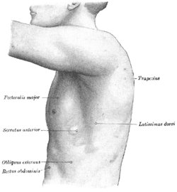
In most cases of winged scapula, damage to the serratus anterior muscle causes the deformation of the back. The serratus anterior muscle attaches to the medial anterior aspect of the scapula (i.e. it attaches on the side closest to the spine and runs along the side of the scapula that faces the ribcage) and normally anchors the scapula against the rib cage. When the serratus anterior contracts, upward rotation, abduction, and weak elevation of the scapula occurs, allowing the arm to be raised above the head. [4] The long thoracic nerve innervates the serratus anterior; therefore, damage to or impingement of this nerve can result in weakening or paralysis of the muscle. [5] If this occurs, the scapula may slip away from the rib cage, giving it the wing-like appearance on the upper back. This characteristic may particularly be seen when the affected person pushes against resistance. The person may also have limited ability to lift their arm above their head.

In FSHD, the winged scapula is detected during contraction of the glenohumeral joint. In this movement, the glenohumeral joint atypically and concurrently abducts and the scapula internally rotates. [3]

Causes

Winging of the scapula is divided into two categories, medial and lateral, according to the direction of winging. [6] Medial winging is more common, being caused by serratus anterior paralysis. [1] This is typically due to damage (i.e. lesions) of the long thoracic nerve. [1] [7] This nerve supplies the serratus anterior, which is located on the side of the thorax and acts to pull the scapula forward. Serratus anterior palsy is a dysfunction that is characteristic of traumatic, non-traumatic, and idiopathic injury to the long thoracic nerve. [1] The second category is the lateral winging which is caused by injury of the spinal accessory nerve. Severe atrophy of the trapezius is seen with accidental damage to the spinal accessory nerve during lymph node biopsy of the neck. There are numerous ways in which the long thoracic nerve can sustain trauma-induced injury. These include, but are not limited to, blunt trauma (e.g. blow to the neck or shoulder, sudden depression of the shoulder girdle, unusual twisting of the neck and shoulder), repetitive movements (as observed in athletic activities such as weight lifting or sports that involve throwing), excessive compression of the shoulder area by straps (see backpack palsy), and various household activities (e.g. gardening, digging, car washing, prolonged abduction of the arms when sleeping, propping up the head to read, etc.). Sometimes, other structures in the body such as inflamed and enlarged subcoracoid or subscapular bursa press on the nerve. Clinical treatments may also cause injury to the long thoracic nerve (iatrogenesis from forceful manipulation, mastectomies with axillary node dissection, surgical treatment of spontaneous pneumothorax, post-general anesthesia for various clinical reasons, and electrical shock, amongst others). [1]

Non-traumatic induced injury to the long thoracic nerve includes, but is not limited to, causes such as viral illness (e.g. influenza, tonsillitis-bronchitis, polio), allergic-drug reactions, drug overdose, toxic exposure (e.g. herbicides, tetanus), C7 radiculopathy, and coarctation of the aorta. [1]

Secondary to serratus anterior palsy, a winged scapula is also caused by trapezius and rhomboid muscle palsy involving the accessory nerve and the dorsal scapular nerve, respectively. [1]

Though the most common causes of a winged scapula is due to serratus anterior palsy, and less commonly trapezius and rhomboid palsy, there are still other circumstances that present the ailment. These incidences include direct injuries to the scapulothoracic muscles (i.e. trapezius and rhomboid muscles), and structural abnormalities (e.g. rotator cuff pathology, shoulder instability, etc.). [1] [8]

Diagnosis

In addition to history and exam, it has been recommended to perform projectional radiography of the neck, chest, shoulder, and thoracic inlet to rule out structural abnormalities such as malunited or greenstick fractures. [1] Computed tomography (CT) or magnetic resonance imaging (MRI) are rarely indicated, but may be useful to rule out certain diagnoses if suspected, such as neurofibromatosis-related injury, intervertebral disc disorder, radiculopathy, and tumors. [1]

Treatment

There are a variety of classifications for winged scapula, and likewise several treatment options. Strength training, particularly of the serratus anterior, may be recommended as this muscle is responsible for holding the medial scapula close to the rib cage. In more severe cases, physical therapy can help by strengthening related muscles. Physical therapy constitutes treatment options if there is weakness of the glenohumeral joint muscles, but if the muscles do not contract clinically and symptoms continue to be severe for more than 3–6 months, surgery may be the next choice. [9] [10]

Physical therapy for a winged scapula will usually incorporate exercises aimed at strengthening the serratus anterior. The push-up plus (PUP) exercise is one of the most commonly prescribed for strengthening the serratus anterior. The push-up plus is usually done in either a push-up position either against a wall or progressed to the floor. Full scapular protraction (the plus) is added after full elbow extension at the end of the usual push-up exercise. The plus phase during the PUP exercise has been shown to elicit the highest average serratus anterior electromyographic (EMG) activity when compared with other SA-activating and closed kinetic chain exercises. [11]

Scapula-to-scapula scapulopexy, pre- and post-operation in person with FSHD. The scapulas are tethered together into a retracted position with an Achilles tendon graft. In the right image, the rhomboid major muscles are distinguishable. Before and after Scapula-to-scapula scapulopexy FSHD.png
Scapula-to-scapula scapulopexy, pre- and post-operation in person with FSHD. The scapulas are tethered together into a retracted position with an Achilles tendon graft. In the right image, the rhomboid major muscles are distinguishable.

Surgical options include neurolysis (chordotomy) and intercostal nerve transfer if a nerve lesion is the cause of winging. For scapular winging not amenable to nerve repair, tendon transfer is an option. Pectoralis major transfer can be done for isolated serratus anterior palsy, and an Eden-Lange procedure can be done for isolated trapezius palsy. When tendon transfer is not feasible, such as in the case of muscular dystrophy or multiple muscular deficits, remaining options include scapulothoracic fusion (also known as scapulodesis), which induces bony fusion between the scapula and the rib cage, and scapulothoracic fixation without arthrodesis (scapulopexy). [3] [10] Even though scapulothoracic fusion has been shown to have successful outcomes, complications were present in over 40% of the 130 patients observed by Kord et al. [12]

Epidemiology

A winged scapula due to serratus anterior palsy is rare. In one report (Fardin et al.), there was an incidence of 15 cases out of 7,000 patients seen in the electromyographical laboratory. In another report (Overpeck and Ghormley), there was only one case out of 38,500 patients observed at the Mayo Clinic. In yet another report (Remak), there were three diagnoses of serratus anterior paralysis throughout a series of 12,000 neurological examinations. [1]

Related Research Articles

<span class="mw-page-title-main">Trapezius</span> Muscle between the lower spine and the shoulder blade

The trapezius is a large paired trapezoid-shaped surface muscle that extends longitudinally from the occipital bone to the lower thoracic vertebrae of the spine and laterally to the spine of the scapula. It moves the scapula and supports the arm.

<span class="mw-page-title-main">Scapula</span> Bone that connects the humerus (upper arm bone) with the clavicle (collar bone)

The scapula, also known as the shoulder blade, is the bone that connects the humerus with the clavicle. Like their connected bones, the scapulae are paired, with each scapula on either side of the body being roughly a mirror image of the other. The name derives from the Classical Latin word for trowel or small shovel, which it was thought to resemble.

<span class="mw-page-title-main">Latissimus dorsi muscle</span> Large, flat back muscle

The latissimus dorsi is a large, flat muscle on the back that stretches to the sides, behind the arm, and is partly covered by the trapezius on the back near the midline. The word latissimus dorsi comes from Latin and means "broadest [muscle] of the back", from "latissimus" ' and "dorsum". The pair of muscles are commonly known as "lats", especially among bodybuilders. The latissimus dorsi is the largest muscle in the upper body.

<span class="mw-page-title-main">Brachial plexus</span> Network of nerves

The brachial plexus is a network of nerves formed by the anterior rami of the lower four cervical nerves and first thoracic nerve. This plexus extends from the spinal cord, through the cervicoaxillary canal in the neck, over the first rib, and into the armpit, it supplies afferent and efferent nerve fibers to the chest, shoulder, arm, forearm, and hand.

<span class="mw-page-title-main">Accessory nerve</span> Cranial nerve XI, for head and shoulder movements

The accessory nerve, also known as the eleventh cranial nerve, cranial nerve XI, or simply CN XI, is a cranial nerve that supplies the sternocleidomastoid and trapezius muscles. It is classified as the eleventh of twelve pairs of cranial nerves because part of it was formerly believed to originate in the brain. The sternocleidomastoid muscle tilts and rotates the head, whereas the trapezius muscle, connecting to the scapula, acts to shrug the shoulder.

<span class="mw-page-title-main">Long thoracic nerve</span> Large nerve

The long thoracic nerve is a branch of the brachial plexus derived from cervical nerves C5-C7 that innervates the serratus anterior muscle.

<span class="mw-page-title-main">Deltoid muscle</span> Shoulder muscle

The deltoid muscle is the muscle forming the rounded contour of the human shoulder. It is also known as the 'common shoulder muscle', particularly in other animals such as the domestic cat. Anatomically, the deltoid muscle appears to be made up of three distinct sets of muscle fibers, namely the

  1. anterior or clavicular part
  2. posterior or scapular part
  3. intermediate or acromial part
<span class="mw-page-title-main">Upper limb</span> Consists of the arm, forearm, and hand

The upper limbs or upper extremities are the forelimbs of an upright-postured tetrapod vertebrate, extending from the scapulae and clavicles down to and including the digits, including all the musculatures and ligaments involved with the shoulder, elbow, wrist and knuckle joints. In humans, each upper limb is divided into the arm, forearm and hand, and is primarily used for climbing, lifting and manipulating objects.

<span class="mw-page-title-main">Levator scapulae muscle</span> Slender skeletal muscle at the back and side of the neck

The levator scapulae is a slender skeletal muscle situated at the back and side of the neck. It originates from the transverse processes of the four uppermost cervical vertebrae; it inserts onto the upper portion of the medial border of the scapula. It is innervated by the cervical nerves C3-C4, and frequently also by the dorsal scapular nerve. As the Latin name suggests, its main function is to lift the scapula.

<span class="mw-page-title-main">Rhomboid major muscle</span> Skeletal muscle in the human back

The rhomboid major is a skeletal muscle of the back that connects the scapula with the vertebrae of the spinal column. It originates from the spinous processes of the thoracic vertebrae T2-T5 and supraspinous ligament; it inserts onto the lower portion of the medial border of the scapula. It acts together with the rhomboid minor to keep the scapula pressed against thoracic wall and to retract the scapula toward the vertebral column.

<span class="mw-page-title-main">Rhomboid minor muscle</span> Skeletal muscle of the upper back

In human anatomy, the rhomboid minor is a small skeletal muscle of the back that connects the scapula to the vertebrae of the spinal column. It arises from the nuchal ligament, and the 7th cervical and 1st thoracic vertebrae and intervening supraspinous ligaments; it inserts onto the medial border of the scapula. It is innervated by the dorsal scapular nerve.

<span class="mw-page-title-main">Serratus anterior muscle</span> Muscle on the surface of the ribs

The serratus anterior is a muscle of the chest. It originates at the side of the chest from the upper 8 or 9 ribs; it inserts along the entire length of the anterior aspect of the medial border of the scapula. It is innervated by the long thoracic nerve from the brachial plexus. The serratus anterior acts to pull the scapula forward around the thorax.

<span class="mw-page-title-main">Facioscapulohumeral muscular dystrophy</span> Medical condition

Facioscapulohumeral muscular dystrophy (FSHD) is a type of muscular dystrophy, a group of heritable diseases that cause degeneration of muscle and progressive weakness. Per the name, FSHD tends to sequentially weaken the muscles of the face, those that position the scapula, and those overlying the humerus bone of the upper arm. These areas can be spared, and muscles of other areas usually are affected, especially those of the chest, spine, abdomen, and shin. Almost any skeletal muscle can be affected in severe disease. Abnormally positioned, or winged, scapulas are common, as is the inability to lift the foot, known as foot drop. The two sides of the body are often affected unequally. Weakness typically manifests at ages 15 – 30 years. FSHD can also cause hearing loss and blood vessel abnormalities in the back of the eye.

<span class="mw-page-title-main">Shoulder joint</span> Synovial ball and socket joint in the shoulder

The shoulder joint is structurally classified as a synovial ball-and-socket joint and functionally as a diarthrosis and multiaxial joint. It involves an articulation between the glenoid fossa of the scapula and the head of the humerus. Due to the very loose joint capsule that gives a limited interface of the humerus and scapula, it is the most mobile joint of the human body.

<span class="mw-page-title-main">Shoulder girdle</span> Set of bones which connects the arm to the axial skeleton on each side

The shoulder girdle or pectoral girdle is the set of bones in the appendicular skeleton which connects to the arm on each side. In humans it consists of the clavicle and scapula; in those species with three bones in the shoulder, it consists of the clavicle, scapula, and coracoid. Some mammalian species have only the scapula.

<span class="mw-page-title-main">Spine of scapula</span> Bony plate on the scapula

The spine of the scapula or scapular spine is a prominent plate of bone, which crosses obliquely the medial four-fifths of the scapula at its upper part, and separates the supra- from the infraspinatous fossa.

Shoulder surgery is a means of treating injured shoulders. Many surgeries have been developed to repair the muscles, connective tissue, or damaged joints that can arise from traumatic or overuse injuries to the shoulder.

<span class="mw-page-title-main">Accessory nerve disorder</span> Disorder caused due to injury to the spinal accessory nerve (11th cranial nerve or Cranial Nerve XI)

Accessory nerve disorder is an injury to the spinal accessory nerve which results in diminished or absent function of the sternocleidomastoid muscle and upper portion of the trapezius muscle.

<span class="mw-page-title-main">Shoulder impingement syndrome</span> Medical condition

Shoulder impingement syndrome is a syndrome involving tendonitis of the rotator cuff muscles as they pass through the subacromial space, the passage beneath the acromion. It is particularly associated with tendonitis of the supraspinatus muscle. This can result in pain, weakness, and loss of movement at the shoulder.

The Eden-Lange procedure is an orthopedic procedure to alleviate the symptoms of trapezius palsy when more conservative measures, such as spontaneous resolution and surgical nerve repair are not promising. The rhomboid major, rhomboid minor, and levator scapulae muscles are transferred laterally along the scapula to replace the functions of the lower, middle, and upper fibers of the trapezius, respectively. The transferred muscles hold the scapula in a more medial and upwardly rotated position, without winging.

References

  1. 1 2 3 4 5 6 7 8 9 10 11 Martin RM, Fish DE (March 2008). "Scapular winging: anatomical review, diagnosis, and treatments". Current Reviews in Musculoskeletal Medicine. 1 (1): 1–11. doi:10.1007/s12178-007-9000-5. PMC   2684151 . PMID   19468892.
  2. Lee SG, Kim JH, Lee SY, Choi IS, Moon ES (June 2006). "Winged scapula caused by rhomboideus and trapezius muscles rupture associated with repetitive minor trauma: a case report". Journal of Korean Medical Science. 21 (3): 581–4. doi:10.3346/jkms.2006.21.3.581. PMC   2729973 . PMID   16778411.
  3. 1 2 3 4 Giannini S, Faldini C, Pagkrati S, Grandi G, Digennaro V, Luciani D, Merlini L (October 2007). "Fixation of winged scapula in facioscapulohumeral muscular dystrophy". Clinical Medicine & Research. 5 (3): 155–62. doi:10.3121/cmr.2007.736. PMC   2111408 . PMID   18056023.
  4. "Serratus Anterior". ExRx.net.
  5. Nath RK, Lyons AB, Bietz G (March 2007). "Microneurolysis and decompression of long thoracic nerve injury are effective in reversing scapular winging: long-term results in 50 cases". BMC Musculoskeletal Disorders. 8: 25. doi: 10.1186/1471-2474-8-25 . PMC   1831472 . PMID   17343759.
  6. Cole, Brian J.; Sekiya, Jon K. (2013). Surgical Techniques of the Shoulder, Elbow and Knee in Sports Medicine E-Book: Expert Consult - Online and Print. Elsevier Health Sciences. p. 334. ISBN   9781455723584.
  7. Le Nail LR, Bacle G, Marteau E, Corcia P, Favard L, Laulan J (June 2014). "Isolated paralysis of the serratus anterior muscle: surgical release of the distal segment of the long thoracic nerve in 52 patients". Orthopaedics & Traumatology, Surgery & Research. 100 (4 Suppl): S243-8. doi: 10.1016/j.otsr.2014.03.004 . PMID   24703793.
  8. Schmitz C, Sodian R, Witt TN, Juchem G, Lang N, Bruegger C, et al. (April 2009). "Winged scapula after aortic valve replacement". The Annals of Thoracic Surgery. 87 (4): 1277–9. doi:10.1016/j.athoracsur.2008.08.050. PMID   19324173.
  9. Galano GJ, Bigliani LU, Ahmad CS, Levine WN (March 2008). "Surgical treatment of winged scapula". Clinical Orthopaedics and Related Research. 466 (3): 652–60. doi:10.1007/s11999-007-0086-2. PMC   2505206 . PMID   18196359.
  10. 1 2 "Scapular Winging". NYU Medical Center. Archived from the original on September 29, 2012.
  11. Kang FJ, Ou HL, Lin KY, Lin JJ (November 2019). "Serratus Anterior and Upper Trapezius Electromyographic Analysis of the Push-Up Plus Exercise: A Systematic Review and Meta-Analysis". Journal of Athletic Training. 54 (11): 1156–1164. doi:10.4085/1062-6050-237-18. PMC   6863690 . PMID   31584855.
  12. Kord D, Liu E, Horner NS, Athwal GS, Khan M, Alolabi B (April 2020). "Outcomes of scapulothoracic fusion in facioscapulohumeral muscular dystrophy: A systematic review". Shoulder & Elbow. 12 (2): 75–90. doi:10.1177/1758573219866195. PMC   7153204 . PMID   32313557.