Tarsal tunnel syndrome

Last updated
Tarsal tunnel syndrome
Other namesPosterior tibial neuralgia
Gray442.png
The mucous sheaths of the tendons around the ankle. Medial aspect.
Specialty Neurology   OOjs UI icon edit-ltr-progressive.svg

Tarsal tunnel syndrome (TTS) is a nerve entrapment syndrome causing a painful foot condition in which the tibial nerve is compressed as it travels through the tarsal tunnel. [1] [2] This tunnel is found along the inner leg behind the medial malleolus (bump on the inside of the ankle). The posterior tibial artery, tibial nerve, and tendons of the tibialis posterior, flexor digitorum longus, and flexor hallucis longus muscles travel in a bundle through the tarsal tunnel. Inside the tunnel, the nerve splits into three segments. One nerve (calcaneal) continues to the heel, the other two (medial and lateral plantar nerves) continue on to the bottom of the foot. The tarsal tunnel is delineated by bone on the inside and the flexor retinaculum on the outside.

Contents

Patients with TTS typically complain of numbness in the foot radiating to the big toe and the first three toes, pain, burning, electrical sensations, and tingling over the base of the foot and the heel. [1] Depending on the area of entrapment, other areas can be affected. If the entrapment is high, the entire foot can be affected as varying branches of the tibial nerve can become involved. Ankle pain is also present in patients who have high level entrapments. Inflammation or swelling can occur within this tunnel for a number of reasons. The flexor retinaculum has a limited ability to stretch, so increased pressure will eventually cause compression on the nerve within the tunnel. As pressure increases on the nerves, the blood flow decreases. [1] Nerves respond with altered sensations like tingling and numbness. Fluid collects in the foot when standing and walking and this makes the condition worse. As small muscles lose their nerve supply they can create a cramping feeling.

Symptoms

Some of the symptoms are:

Tinel's sign is a tingling electric shock sensation that occurs when you tap over an affected nerve. The sensation usually travels into the foot but can also travel up the inner leg as well.

Causes

3D still showing tarsal tunnel syndrome Tarsal Tunnel.jpg
3D still showing tarsal tunnel syndrome

It is difficult to determine the exact cause of tarsal tunnel syndrome. It is important to attempt to determine the source of the problem. Treatment and the potential outcome of the treatment may depend on the cause. Anything that creates pressure in the tarsal tunnel can cause TTS. This would include benign tumors or cysts, bone spurs, inflammation of the tendon sheath, nerve ganglions, or swelling from a broken or sprained ankle. Varicose veins (that may or may not be visible) can also cause compression of the nerve. TTS is more common in athletes and other active people. These people put more stress on the tarsal tunnel area. Flat feet may cause an increase in pressure in the tunnel region and this can cause nerve compression. Those with lower back problems may have symptoms. Back problems with the L4, L5 and S1 regions are suspect and might suggest a "Double Crush" issue: one "crush" (nerve pinch or entrapment) in the lower back, and the second in the tunnel area. In some cases, TTS can simply be idiopathic. [1]

This is an image of a normal arched foot. Normal Arch.jpg
This is an image of a normal arched foot.
When comparing to the normal arch image, this image of fallen arches helps create a visualization of how the tibial nerve can be strained and compressed due to the curvature. Fallen arch (flat feet).jpg
When comparing to the normal arch image, this image of fallen arches helps create a visualization of how the tibial nerve can be strained and compressed due to the curvature.

Rheumatoid Arthritis has also been associated with TTS. [3]

Neurofibromatosis can also cause TTS. This is a disease that results in the formation of pigmented, cutaneous neurofibromas. These masses, in a specific case, were shown to have the ability to invade the tarsal tunnel causing pressure, therefore resulting in TTS. [4]

Diabetes makes the peripheral nerve susceptible to nerve compression, as part of the double crush hypothesis. [5] In contrast to carpal tunnel syndrome due to one tunnel at the wrist for the median nerve, there are four tunnels in the medial ankle for tarsal tunnels syndrome. [6] If there is a positive Tinel sign when you tap over the inside of the ankle, such that tingling is felt into the foot, then there is an 80% chance that decompressing the tarsal tunnel will relieve the symptoms of pain and numbness in a diabetic with tarsal tunnel syndrome. [7]

Risk factors

Anything compromising the tunnel of the posterior tibial nerve proves significant in the risk of causing TTS. Neuropathy can occur in the lower limb through many modalities, some of which include obesity and inflammation around the joints. By association, this includes risk factors such as RA, compressed shoes, pregnancy, diabetes and thyroid diseases [8]

Diagnosis

Diagnosis is based upon physical examination findings. Patients' pain history and a positive Tinel's sign are the first steps in evaluating the possibility of tarsal tunnel syndrome. X-ray can rule out fracture. MRI can assess for space occupying lesions or other causes of nerve compression. Ultrasound can assess for synovitis or ganglia. Nerve conduction studies alone are not, but they may be used to confirm the suspected clinical diagnosis. Common causes include trauma, varicose veins, neuropathy and space-occupying anomalies within the tarsal tunnel. Tarsal tunnel syndrome is also known to affect both athletes and individuals that stand a lot. [1]

A neurologist or a physiatrist usually administers nerve conduction tests or supervises a trained technologist. During this test, electrodes are placed at various spots along the nerves in the legs and feet. Both sensory and motor nerves are tested at different locations. Electrical impulses are sent through the nerve and the speed and intensity at which they travel is measured. If there is compression in the tunnel, this can be confirmed and pinpointed with this test. Some doctors do not feel that this test is necessarily a reliable way to rule out TTS. [1] Some research indicates that nerve conduction tests will be normal in at least 50% of the cases.

Given the unclear role of electrodiagnostics in the diagnosis of tarsal tunnel syndrome, efforts have been made in the medical literature to determine which nerve conduction studies are most sensitive and specific for tibial mononeuropathy at the level of the tarsal tunnel. An evidence-based practice topic put forth by the professional organization, the American Association of Neuromuscular & Electrodiagnostic Medicine has determined that Level C, Class III evidence exists for the use of tibial motor nerve conduction studies, medial and lateral plantar mixed nerve conduction studies, and medial and lateral plantar sensory nerve conduction studies. The role of needle electromyography remains less defined. [9]

Tarsal tunnel syndrome (TTS) is most closely related to carpal tunnel syndrome (CTS), but is much less common. [10] Studies have found that patients with rheumatoid arthritis (RA) show signs of distal limb neuropathy. The posterior tibial nerve serves victim to peripheral neuropathy and often show signs of TTS amongst RA patients. Therefore, TTS is a common discovery found in the autoimmune disorder of rheumatoid arthritis [11]

Increased pressure and high loads on the ankle joint can cause TTS, as can smaller than normal shoes. In this picture, most of the load is placed upon the knee and ankle joint. Increased ankle loads TTS.jpg
Increased pressure and high loads on the ankle joint can cause TTS, as can smaller than normal shoes. In this picture, most of the load is placed upon the knee and ankle joint.

Prevention

The exact cause of tarsal tunnel syndrome (TTS) can vary from patient to patient. However the same result is true for all patients, the compression of the posterior tibial nerve and its branches as it travels around the medial malleolus causes pain and irritation for the patient. [12] There are many possible causes for compression of the tibial nerve therefore there are a variety of prevention strategies. One being immobilization, by placing the foot in a neutral position with a brace, pressure is relieved from the tibial nerve thus reducing patients pain. [13] [14] [15] Eversion, inversion, and plantarflexion all can cause compression of the tibial nerve therefore in the neutral position the tibial nerve is less agitated. Typically this is recommended for the patient to do while sleeping. Another common problem is improper footwear, having shoes deforming the foot due to being too tight can lead to increased pressure on the tibial nerve. [12] Having footwear that tightens the foot for extended periods of time even will lead to TTS. Therefore, by simply having properly fitted shoes TTS can be prevented.

Treatment

Treatments typically include rest, manipulation, strengthening of tibialis anterior, tibialis posterior, peroneus and short toe flexors, casting with a walker boot, corticosteroid and anesthetic injections, hot wax baths, wrapping, compression hose, and orthotics. Medications may include various anti-inflammatories such as Anaprox, or other medications such as Ultracet, Neurontin and Lyrica. Lidocaine patches are also a treatment that helps some patients.

Conservative treatment (nonsurgical)

There are multiple ways that tarsal tunnel can be treated and the pain can be reduced. The initial treatment, whether it be conservative or surgical, depends on the severity of the tarsal tunnel and how much pain the patient is in. There was a study done that treated patients diagnosed with tarsal tunnel syndrome with a conservative approach. Meaning that the program these patients were participated in consisted of physiotherapy exercises and orthopedic shoe inserts in addition to that program. There were fourteen patients that had supplementary tibial nerve mobilization exercises. They were instructed to sit on the edge of a table in a slumped position, have their ankle taken into dorsiflexion and ankle eversion then the knee was extended and flexed to obtain the optimal tibial nerve mobilization. Patients in both groups showed positive progress from both programs. [16] The medial calcaneal, medial plantar and lateral plantar nerve areas all had a reduction in pain after successful nonoperative or conservative treatment. [17] There is also the option of localized steroid or cortisone injection that may reduce the inflammation in the area, therefore relieving pain. Or just a simple reduction in the patient's weight to reduce the pressure in the area. [18]

Tarsal tunnel release Tarsal Tunnel Decompression.png
Tarsal tunnel release

Surgical treatment

If non-invasive treatment measures fail, tarsal tunnel release surgery may be recommended to decompress the area. The incision is made behind the ankle bone and then down towards but not as far as the bottom of foot. The posterior tibial nerve is identified above the ankle. It is separated from the accompanying artery and vein and then followed into the tunnel. The nerves are released. Cysts or other space-occupying problems may be corrected at this time. If there is scarring within the nerve or branches, this is relieved by internal neurolysis. Neurolysis is when the outer layer of nerve wrapping is opened and the scar tissue is removed from within nerve. Following surgery, a large bulky cotton wrapping immobilizes the ankle joint without plaster. The dressing may be removed at the one-week point and sutures at about three weeks.

Complications may include bleeding, infection, and unpredictable healing. The incision may open from swelling. There may be considerable pain and cramping. Regenerating nerve fibers may create shooting pains. Patients may have hot or cold sensations and may feel worse than before surgery. Crutches are usually recommended for the first two weeks, as well as elevation to minimize swelling. The nerve will grow at about one inch per month. One can expect to continue the healing process over the course of about one year.

Many patients report good results. Some, however, experience no improvement or a worsening of symptoms. In the Pfeiffer article (Los Angeles, 1996),[ full citation needed ] fewer than 50% of the patients reported improvement, and there was a 13% complication rate.

Tarsal tunnel can greatly impact patients' quality of life. Depending on the severity, the ability to walk distances people normally take for granted (such as grocery shopping) may become compromised. Proper pain management and counseling is often required.

Results of surgery can be maximized if all four of the medial ankle tunnels are released and you walk with a walker the day after surgery. Success can be improved to 80%. [19]

Incidence

Though TTS is rare, its cause can be determined in 70% of reported cases. In the workplace TTS is considered a musculoskeletal disorder and accounts for 1.8 million cases a year, which accumulates to about $15–$20 billion a year [20] New studies indicate an occurrence of TTS in sports placing high loads on the ankle joint (3). This can be seen in figure 1. TTS occurs more dominantly in active adults, with a higher pervasiveness among women. Active adults that experience more jumping and landing on the ankle joint are more susceptible (see figure 2). Though athletics and sport are correlations, cases are individualistically assessed because of the oddity.

Athletic activities

The athletic population tends to put themselves at greater risk of TTS due to the participation in sports that involve the lower extremities. Strenuous activities involved in athletic activities put extra strain on the ankle and therefore can lead to the compression of the tibial nerve. [21] Activities that especially involve sprinting and jumping have a greater risk of developing TTS. This is due to the ankle being put in eversion, inversion, and plantarflexion at high velocities. Examples of sports that can lead to TTS include basketball, track, soccer, lacrosse, snowboarding, and volleyball. [22] Participation in these sports should be done cautiously due to the high risk of developing TTS. However athletes will tend to continue to participate in these activities therefore proper stretching, especially in lower extremities, prior to participation can assist in the prevention of developing TTS.

Placing the foot in eversion (top left), inversion (bottom left), or plantar flexion(right) all put strain on the tibial nerve. These positions should be avoided to prevent the development of TTS and can be done so by using a brace to place the foot in a neutral position. Tarsal Tunnel Syndrome Foot Positions.jpg
Placing the foot in eversion (top left), inversion (bottom left), or plantar flexion(right) all put strain on the tibial nerve. These positions should be avoided to prevent the development of TTS and can be done so by using a brace to place the foot in a neutral position.

Famous case

According to South Korea's National Intelligence Service, North Korean leader Kim Jong Un had surgery to correct TTS in his right ankle, the source of a pronounced limp. Kim's disappearance from public for six weeks around the suspected surgery created worldwide speculation about the future of Kim and North Korea. [23]

Society

As stated earlier, musculoskeletal disorders can cost up to $15–$20 billion in direct costs or $45–$55 billion in indirect expenses. This is about $135 million a day. [20] Tests that confirm or correct TTS require expensive treatment options like X-rays, CT-scans, MRI and surgery. The three former options for TTS detect and locate, while the latter is a form of treatment to decompress tibial nerve pressure. [24] Since surgery is the most common form of TTS treatment, high financial burden is placed upon those diagnosed with the rare syndrome.

See also

Related Research Articles

<span class="mw-page-title-main">Carpal tunnel syndrome</span> Medical condition

Carpal tunnel syndrome (CTS) is the collection of symptoms and signs associated with median neuropathy at the carpal tunnel. Most CTS is related to idiopathic compression of the median nerve as it travels through the wrist at the carpal tunnel (IMNCT). Idiopathic means that there is no other disease process contributing to pressure on the nerve. As with most structural issues, it occurs in both hands, and the strongest risk factor is genetics.

Pudendal nerve entrapment (PNE), also known as Alcock canal syndrome, is an uncommon source of chronic pain in which the pudendal nerve is entrapped or compressed in Alcock's canal. There are several different types of PNE based on the site of entrapment anatomically. Pain is positional and is worsened by sitting. Other symptoms include genital numbness, fecal incontinence and urinary incontinence.

<span class="mw-page-title-main">Peroneal nerve paralysis</span> Medical condition

Peroneal nerve paralysis is a paralysis on common fibular nerve that affects patient’s ability to lift the foot at the ankle. The condition was named after Friedrich Albert von Zenker. Peroneal nerve paralysis usually leads to neuromuscular disorder, peroneal nerve injury, or foot drop which can be symptoms of more serious disorders such as nerve compression. The origin of peroneal nerve palsy has been reported to be associated with musculoskeletal injury or isolated nerve traction and compression. Also it has been reported to be mass lesions and metabolic syndromes. Peroneal nerve is most commonly interrupted at the knee and possibly at the joint of hip and ankle. Most studies reported that about 30% of peroneal nerve palsy is followed from knee dislocations.

<span class="mw-page-title-main">Tinel's sign</span> Medical condition

Tinel's sign is a way to detect irritated nerves. It is performed by lightly tapping (percussing) over the nerve to elicit a sensation of tingling or "pins and needles" in the distribution of the nerve. Percussion is usually performed moving distal to proximal. It is named after Jules Tinel.
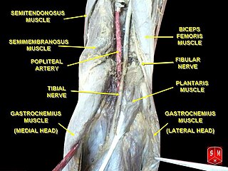
The posterior tibial artery of the lower limb is an artery that carries blood to the posterior compartment of the leg and plantar surface of the foot. It branches from the popliteal artery via the tibial-fibular trunk.

Meralgia paresthetica or meralgia paraesthetica is numbness or pain in the outer thigh not caused by injury to the thigh, but by injury to a nerve that extends from the spinal column to the thigh.

<span class="mw-page-title-main">Flexor digitorum longus muscle</span> Muscle located on the tibial side of the leg

The flexor digitorum longus muscle is situated on the tibial side of the leg. At its origin it is thin and pointed, but it gradually increases in size as it descends. It serves to flex the second, third, fourth, and fifth toes.

The common fibular nerve is a nerve in the lower leg that provides sensation over the posterolateral part of the leg and the knee joint. It divides at the knee into two terminal branches: the superficial fibular nerve and deep fibular nerve, which innervate the muscles of the lateral and anterior compartments of the leg respectively. When the common fibular nerve is damaged or compressed, foot drop can ensue.

The flexor retinaculum of foot is a strong fibrous band in the foot.

<span class="mw-page-title-main">Popliteal artery entrapment syndrome</span> Abnormal popliteal artery compression by surrounding structures

The popliteal artery entrapment syndrome (PAES) is an uncommon pathology that occurs when the popliteal artery is compressed by the surrounding popliteal fossa myofascial structures. This results in claudication and chronic leg ischemia. This condition mainly occurs more in young athletes than in the elderlies. Elderlies, who present with similar symptoms, are more likely to be diagnosed with peripheral artery disease with associated atherosclerosis. Patients with PAES mainly present with intermittent feet and calf pain associated with exercises and relieved with rest. PAES can be diagnosed with a combination of medical history, physical examination, and advanced imaging modalities such as duplex ultrasound, computer tomography, or magnetic resonance angiography. Management can range from non-intervention to open surgical decompression with a generally good prognosis. Complications of untreated PAES can include stenotic artery degeneration, complete popliteal artery occlusion, distal arterial thromboembolism, or even formation of an aneurysm.

<span class="mw-page-title-main">Tarsal tunnel</span> Canal in the ankle area

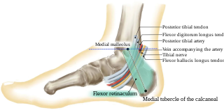
The tarsal tunnel is a passage found along the inner leg underneath the medial malleolus of the ankle.

<span class="mw-page-title-main">Superior cluneal nerves</span>

The superior cluneal nerves are pure sensory nerves that innervate the skin of the upper part of the buttocks. They are the terminal ends of the L1-L3 spinal nerve dorsal rami lateral branches. They are one of three different types of cluneal nerves. They travel inferiorly through multiple layers of muscles, then traverse osteofibrous tunnels between the thoracolumbar fascia and iliac crest.

<span class="mw-page-title-main">Ulnar nerve entrapment</span> Medical condition

Ulnar nerve entrapment is a condition where pressure on the ulnar nerve as it passes through the cubital tunnel causes nerve dysfunction (neuropathy). The symptoms of neuropathy are paresthesia (tingling) and numbness primarily affecting the little finger and ring finger of the hand. Ulnar neuropathy can progress to weakness and atrophy of the muscles in the hand. Symptoms can be alleviated by attempts to keep the elbow from flexing while sleeping, such as sticking one’s arm in the pillow case, so the pillow restricts flexion.

<span class="mw-page-title-main">Ulnar neuropathy</span> Medical condition

Ulnar neuropathy is a disorder involving the ulnar nerve. Ulnar neuropathy may be caused by entrapment of the ulnar nerve with resultant numbness and tingling. It may also cause weakness or paralysis of the muscles supplied by the nerve.

Anterior interosseous syndrome is a medical condition in which damage to the anterior interosseous nerve (AIN), a distal motor and sensory branch of the median nerve, classically with severe weakness of the pincer movement of the thumb and index finger, and can cause transient pain in the wrist.

<span class="mw-page-title-main">Pronator teres syndrome</span> Medical condition

Pronator teres syndrome is a compression neuropathy of the median nerve at the elbow. It is rare compared to compression at the wrist or isolated injury of the anterior interosseous branch of the median nerve.

<span class="mw-page-title-main">Nerve compression syndrome</span> Human disease

Nerve compression syndrome, or compression neuropathy, or nerve entrapment syndrome, is a medical condition caused by chronic, direct pressure on a peripheral nerve. It is known colloquially as a trapped nerve, though this may also refer to nerve root compression. Its symptoms include pain, tingling, numbness and muscle weakness. The symptoms affect just one particular part of the body, depending on which nerve is affected. The diagnosis is largely clinical and can be confirmed with diagnostic nerve blocks. Occasionally imaging and electrophysiology studies aid in the diagnosis. Timely diagnosis is important as untreated chronic nerve compression may cause permanent damage. A surgical nerve decompression can relieve pressure on the nerve but cannot always reverse the physiological changes that occurred before treatment. Nerve injury by a single episode of physical trauma is in one sense an acute compression neuropathy but is not usually included under this heading, as chronic compression takes a unique pathophysiological course.

<span class="mw-page-title-main">Median nerve palsy</span> Medical condition

Injuries to the arm, forearm or wrist area can lead to various nerve disorders. One such disorder is median nerve palsy. The median nerve controls the majority of the muscles in the forearm. It controls abduction of the thumb, flexion of hand at wrist, flexion of digital phalanx of the fingers, is the sensory nerve for the first three fingers, etc. Because of this major role of the median nerve, it is also called the eye of the hand. If the median nerve is damaged, the ability to abduct and oppose the thumb may be lost due to paralysis of the thenar muscles. Various other symptoms can occur which may be repaired through surgery and tendon transfers. Tendon transfers have been very successful in restoring motor function and improving functional outcomes in patients with median nerve palsy.

Femoral nerve dysfunction, also known as femoral neuropathy, is a rare type of peripheral nervous system disorder that arises from damage to nerves, specifically the femoral nerve. Given the location of the femoral nerve, indications of dysfunction are centered around the lack of mobility and sensation in lower parts of the legs. The causes of such neuropathy can stem from both direct and indirect injuries, pressures and diseases. Physical examinations are usually first carried out, depending on the high severity of the injury. In the cases of patients with hemorrhage, imaging techniques are used before any physical examination. Another diagnostic method, electrodiagnostic studies, are recognized as the gold standard that is used to confirm the injury of the femoral nerve. After diagnosis, different treatment methods are provided to the patients depending upon their symptoms in order to effectively target the underlying causes. Currently, femoral neuropathy is highly underdiagnosed and its precedent medical history is not well documented worldwide.

<span class="mw-page-title-main">Deep gluteal syndrome</span> Medical condition

Deep gluteal syndrome describes the non-discogenic extrapelvic entrapment of the sciatic nerve in the deep gluteal space. It is an extension of non-discogenic sciatic nerve entrapment beyond the traditional model of piriformis syndrome. Symptoms are pain or dysthesias the buttocks, hip, and posterior thigh with or without radiating leg pain. Patients often report pain when sitting. The two most common causes are piriformis syndrome and fibrovascular bands, but many other causes exist. Diagnosis is usually done through physical examination, magnetic resonance imaging, magnetic resonance neurography, and diagnostic nerve blocks. Surgical treatment is an endoscopic sciatic nerve decompression.

References

  1. 1 2 3 4 5 6 7 8 Yates, Ben (2009). Merriman's Assessment of the Lower Limb (3rd ed.). New York: Churchill Livingstone. ISBN   978-0-08-045107-7.
  2. "Tarsaltunnelsyndrom". Gesundheitsinfo news. Retrieved 2023-12-16.
  3. Baylan SP, Paik SW, Barnert AL, Ko KH, Yu J, Persellin RH (1981). "Prevalence of the tarsal tunnel syndrome in rheumatoid arthritis". Rheumatol Rehabil. 20 (3): 148–50. doi:10.1093/rheumatology/20.3.148. PMID   7280489.
  4. Mirick, Anika L., Gerald B. Bornstein, and Laura W. Bancroft. "Radiologic Case Study." Orthopedics 36.81 (2013): 154–57. Web. 22 Apr. 2014.
  5. Dellon AL, Mackinnon SE, Seiler WA (1988). "Susceptibility of the diabetic nerve to chronic compression". Ann Plast Surg. 20 (2): 117–119. doi:10.1097/00000637-198802000-00004. PMID   3355055.
  6. Mackinnon SE, Dellon AL (1987). "Homologies between the tarsal and carpal tunnels: Implications for treatment of the tarsal tunnel syndrome". Contemp Orthop. 14: 75–79.
  7. Lee C, Dellon AL (2004). "Prognostic ability of Tinel sign in determining outcome for decompression surgery decompression surgery in diabetic and non-diabetic neuropathy". Ann Plast Surg. 53 (6): 523–27. doi:10.1097/01.sap.0000141379.55618.87. PMID   15602246.
  8. Beltran L. S.; Bencardino J.; Ghazikhanian V.; Beltran J. (2010). "Entrapment Neuropathies III: Lower Limb". Seminars in Musculoskeletal Radiology. 14 (5): 501–511. doi:10.1055/s-0030-1268070. PMID   21072728.
  9. "Usefulness of electrodiagnostic techniques in the evaluation of suspected tarsal tunnel syndrome: An evidence-based review". aanem.org/getmedia/51417557-424c-4c29-be6a-5bbaff64517c/TarsalTunnel.pdf.aspx. Retrieved 15 February 2015.
  10. Ahmad, M. M., Tsang, K. K., Mackenney, P. J., & Adedapo, A. O. (2012). Tarsal tunnel syndrome: A literature review. Foot & Ankle Surgery (Elsevier Science), 18(3), 149–152.
  11. Baylan, S. P., S. W. Paik, A. L. Barnert, K. H. Ko, J. Yu, and R. H. Persellin. "Prevalence Of The Tarsal Tunnel Syndrome In Rheumatoid Arthritis." Rheumatology 20.3 (1981): 148–150.
  12. 1 2 Low, Hu L., and George Stephenson. "These Boots Weren't Made for Walking: Tarsal Tunnel Syndrome." Canadian Medical Association Journal 176.10 (2007): 1415–416.
  13. Gondring, William H., Elly Trepman, and Byron Shields. "Tarsal Tunnel Syndrome: Assessment of Treatment Outcome with an Anatomic Pain Intensity Scale."Foot and Ankle Surgery 15.3 (2009): 133–38.
  14. Bracilovic, A., A. Nihal, V. L. Houston, A. C. Beatle, Z. S. Rosenberg, and E. Trepman. "Effect of Foot and Ankle Position on Tarsal Tunnel Compartment Volume." Foot and Ankle International 27.6 (2006): 421–37.
  15. Nakasa, Tomoyuki, Kohei Fukuhara, Nobuo Adachi, and Mitsuo Ochi. "Painful Os Intermetatarseum in Athletes: Report of Four Cases and Review of the Literature." Archives of Orthopaedic and Trauma Surgery 127.4 (2007): 261–64. Print.
  16. Kavlak Y, Uygur F (2011). "Effects of nerve mobilization exercise as an adjunct to the conservative treatment for patients with tarsal tunnel syndrome". J Manipulative Physiol Ther. 34 (7): 441–8. doi:10.1016/j.jmpt.2011.05.017. PMID   21875518.
  17. Gondring WH1, Trepman E, Shields B. (2008). Tarsal tunnel syndrome: assessment of treatment outcome with an anatomic pain intensity scale. Foot Ankle Surg. 15(3):133-8
  18. Edwards William G.; Lincoln C. Robert; Bassett Frank H.; Goldner J. Leonard (1969). "The Tarsal Tunnel Syndrome Diagnosis and Treatment". JAMA. 207 (4): 716–720. doi:10.1001/jama.1969.03150170042009. PMID   4302951.
  19. Mullick T, Dellon AL. Results of decompression of four medial ankle tunnels in the treatment of tarsal tunnels syndrome. J Reconstr Microsurg. 2008;24:119–126.
  20. 1 2 . Jeffress, Charles N. "Work-related Musculoskeletal Disorders (MSDs)." Work-related Musculoskeletal Disorders (MSDs). Occupational Safety & Health Administration, n.d. Web. 11 May 2014.
  21. Kinoshita, M. "Tarsal Tunnel Syndrome in Athletes." American Journal of Sports Medicine 34.8 (2006): 1307–312.
  22. Ramani, William, David H. Perrin, and Tim Whiteley. "Tarsal Tunnel Syndrome: Case Study of a Male Collegiate Athlete." Journal of Sports Rehabilitation 6 (n.d.): 364-70.
  23. Almasy, KJ Kwon,Steven Almasy,Steve (2014-10-28). "South Korea: Kim Jong Un had ankle surgery to remove cyst". CNN. Retrieved 2021-11-23.{{cite web}}: CS1 maint: multiple names: authors list (link)
  24. Manasseh, N., Cherian, V., & Abel, L. (2009). Malunited calcaneal fracture fragments causing tarsal tunnel syndrome: A rare cause. Foot & Ankle Surgery (Elsevier Science), 15(4), 207–209.