Lateral cutaneous nerve of thigh

Last updated
Lateral cutaneous nerve of thigh
Lumbar plexus.svg
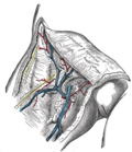
Plan of lumbar plexus. (Lateral femoral cutaneous visible at left.)
Gray825and830.PNG
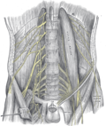
Cutaneous nerves of the right lower extremity. Front and posterior views.
Details
From Lumbar plexus (L2–L3)
InnervatesSkin on the lateral part of the thigh
Identifiers
Latin nervus cutaneus femoris lateralis
TA98 A14.2.07.011
TA2 6521
FMA 16485
Anatomical terms of neuroanatomy

The lateral cutaneous nerve of the thigh (also called the lateral femoral cutaneous nerve) is a cutaneous nerve of the thigh. It originates from the dorsal divisions of the second and third lumbar nerves from the lumbar plexus. It passes under the inguinal ligament to reach the thigh. It supplies sensation to the skin on the lateral part of the thigh by an anterior branch and a posterior branch.

Contents

The lateral cutaneous nerve of the thigh can be investigated using ultrasound. Local anaesthetic can be injected around the nerve for skin grafts and surgery around the outer thigh. Nerve compression (usually around the inguinal ligament) can cause meralgia paraesthetica.

Structure

The nerve is usually 1-2 mm thick. [1]

Origin

The lateral cutaneous nerve of the thigh is a nerve of the lumbar plexus. [2] [1] It arises from the posterior rami of the second and third lumbar nerves (L2-L3). [2] [3]

Course and relations

It passes through psoas major muscle, and emerges from its lateral border. [3] It crosses the iliacus muscle obliquely, toward the anterior superior iliac spine (ASIS). [1] It is crossed by the deep circumflex iliac artery and the deep circumflex iliac vein. [3] It enters the thigh by passing beneath (the lateral part of) the inguinal ligament [4] in the muscular lacuna, [1] or through (the lateral part of) the inguinal ligament itself. [4] It then passes over the sartorius muscle, travelling from medial to lateral. [1]

Branches

The lateral cutaneous nerve of the thigh usually divides into an anterior (or anterolateral) branch and a posterior branch. [3]

Anterior branch

The anterior branch becomes superficial about 10 cm below the inguinal ligament. It divides into branches which are distributed to the skin of the anterior and lateral parts of the thigh, as far down as the knee. [3] The terminal filaments of this nerve frequently communicate with the anterior cutaneous branches of the femoral nerve, and with the infrapatellar branch of the saphenous nerve, forming with them the peripatellar plexus.

Posterior branch

The posterior branch pierces the fascia lata. It subdivides into filaments, which pass backward across the lateral and posterior surfaces of the thigh. It supplies the skin around the greater trochanter. [3]

Distribution

It provides sensory innervation to the lateral aspect of the thigh (as far as the knee), as well as the iliac fascia and perineum of the iliac fascia. [4]

Variation

The lateral cutaneous nerve of the thigh may have multiple branches. [1] Its position with relation to the ASIS can be very variable. [3] It may partially pass through sartorius muscle rather than over its surface. [1] It may be absent, and the sensory supply replaced by branches of the femoral nerve and the ilioinguinal nerve. [1]

Function

The lateral cutaneous nerve of the thigh is a sensory nerve. [2] [1] It supplies the skin on the lateral (outer) part of the thigh. [2] [1]

Clinical significance

Ultrasound

The lateral cutaneous nerve of the thigh can be studied using ultrasound. [1] A patient lies on a bed facing upwards (supine). [3] The ultrasound probe is moved along the length of the nerve, often starting from near the ASIS. [3] The nerve is easier to see over the sartorius muscle than in other subcutaneous tissue, as there is greater contrast. [1] It can sometimes be difficult to see due to surrounding soft tissue. [1]

Nerve block

The lateral cutaneous nerve of the thigh can be blocked with local anaesthetic. [1] Ultrasound is used to guide needle insertion. [3] This is used for procedures in the supplied area of skin, such as surgical incisions over the outer thigh, and skin grafts. [1]

Meralgia paraesthetica

Entrapment of the lateral cutaneous nerve of the thigh is caused by compression of the nerve near the anterior superior iliac spine and the inguinal ligament. [2] This causes meralgia paraesthetica (Bernhardt-Roth syndrome). [2] [5] This may be diagnosed with ultrasound, which changes the morphology of the nerve. [1] Changes can include general enlargement, [1] and a hypoechoic appearance. [3] In patients who only have meralgia paraesthetica on one side, ultrasound scans are performed on both thighs to compare the appearance of the nerve. [3]

History

The lateral cutaneous nerve of the thigh may also be known as the lateral femoral cutaneous nerve. [1]

Additional images

See also

Related Research Articles

<span class="mw-page-title-main">Human leg</span> Lower extremity or limb of the human body (foot, lower leg, thigh and hip)

The leg is the entire lower limb of the human body, including the foot, thigh or sometimes even the hip or buttock region. The major bones of the leg are the femur, tibia, and adjacent fibula. The thigh is between the hip and knee, while the calf (rear) and shin (front) are between the knee and foot.

<span class="mw-page-title-main">Sartorius muscle</span> Longest muscle in the human body

The sartorius muscle is the longest muscle in the human body. It is a long, thin, superficial muscle that runs down the length of the thigh in the anterior compartment.

<span class="mw-page-title-main">Femoral triangle</span> Anatomical region of the thigh

The femoral triangle is an anatomical region of the upper third of the thigh. It is a subfascial space which appears as a triangular depression below the inguinal ligament when the thigh is flexed, abducted and laterally rotated.

<span class="mw-page-title-main">Genitofemoral nerve</span>

The genitofemoral nerve is a mixed branch of the lumbar plexus derived from anterior rami of L1-L2. It splits a genital branch and a femoral branch. It provides sensory innervation to the upper anterior thigh, as well as the skin of the anterior scrotum in males and mons pubis in females. It also provides motor innervation to the cremaster muscle.

Meralgia paresthetica or meralgia paraesthetica is numbness or pain in the outer thigh not caused by injury to the thigh, but by injury to a nerve that extends from the spinal column to the thigh.

<span class="mw-page-title-main">Iliopsoas</span> Joined psoas and the iliacus muscles

The iliopsoas muscle refers to the joined psoas major and the iliacus muscles. The two muscles are separate in the abdomen, but usually merge in the thigh. They are usually given the common name iliopsoas. The iliopsoas muscle joins to the femur at the lesser trochanter. It acts as the strongest flexor of the hip.

<span class="mw-page-title-main">Nerve plexus</span> Network of nerve fibres

A nerve plexus is a plexus of intersecting nerves. A nerve plexus is composed of afferent and efferent fibers that arise from the merging of the anterior rami of spinal nerves and blood vessels. There are five spinal nerve plexuses, except in the thoracic region, as well as other forms of autonomic plexuses, many of which are a part of the enteric nervous system. The nerves that arise from the plexuses have both sensory and motor functions. These functions include muscle contraction, the maintenance of body coordination and control, and the reaction to sensations such as heat, cold, pain, and pressure. There are several plexuses in the body, including:

<span class="mw-page-title-main">Femoral nerve</span> Long nerve down the thigh and inner leg

The femoral nerve is a nerve in the thigh that supplies skin on the upper thigh and inner leg, and the muscles that extend the knee. It is the largest branch of the lumbar plexus.

<span class="mw-page-title-main">Posterior cutaneous nerve of thigh</span> Sensory nerve that supplies the back of the thigh, leg, buttock, and perineum

The posterior cutaneous nerve of the thigh is a sensory nerve of the thigh. It is a branch of the sacral plexus. It supplies the skin of the posterior surface of the thigh, leg, buttock, and also the perineum.

<span class="mw-page-title-main">Lumbar plexus</span> Web of nerves in the lower spine

The lumbar plexus is a web of nerves in the lumbar region of the body which forms part of the larger lumbosacral plexus. It is formed by the divisions of the first four lumbar nerves (L1-L4) and from contributions of the subcostal nerve (T12), which is the last thoracic nerve. Additionally, the ventral rami of the fourth lumbar nerve pass communicating branches, the lumbosacral trunk, to the sacral plexus. The nerves of the lumbar plexus pass in front of the hip joint and mainly support the anterior part of the thigh.

<span class="mw-page-title-main">Iliohypogastric nerve</span>

The iliohypogastric nerve is a nerve that originates from the lumbar plexus that supplies sensation to skin over the lateral gluteal and hypogastric regions and motor to the internal oblique muscles and transverse abdominal muscles.

<span class="mw-page-title-main">Ilioinguinal nerve</span> Branch of the first lumbar nerve

The ilioinguinal nerve is a branch of the first lumbar nerve (L1). It separates from the first lumbar nerve along with the larger iliohypogastric nerve. It emerges from the lateral border of the psoas major just inferior to the iliohypogastric, and passes obliquely across the quadratus lumborum and iliacus. The ilioinguinal nerve then perforates the transversus abdominis near the anterior part of the iliac crest, and communicates with the iliohypogastric nerve between the transversus and the internal oblique muscle.

<span class="mw-page-title-main">Obturator nerve</span>

The obturator nerve in human anatomy arises from the ventral divisions of the second, third, and fourth lumbar nerves in the lumbar plexus; the branch from the third is the largest, while that from the second is often very small.

<span class="mw-page-title-main">Femoral sheath</span> Anatomical structure of the upper thigh

The femoral sheath is a funnel-shaped downward extension of abdominal fascia within which the femoral artery and femoral vein pass between the abdomen and the thigh. The femoral sheath is subdivided by two vertical partitions to form three compartments ; the medial compartment is known as the femoral canal and contains lymphatic vessels and a lymph node, whereas the intermediate canal and the lateral canal accommodate the femoral vein and the femoral artery (respectively). Some neurovascular structures perforate the femoral sheath. Topographically, the femoral sheath is contained within the femoral triangle.

The saphenous nerve is the largest cutaneous branch of the femoral nerve. It is derived from the lumbar plexus (L3-L4). It is a strictly sensory nerve, and has no motor function. It commences in the proximal (upper) thigh and travels along the adductor canal. Upon exiting the adductor canal, the saphenous nerve terminates by splitting into two terminal branches: the sartorial nerve, and the infrapatellar nerve. The saphenous nerve is responsible for providing sensory innervation to the skin of the anteromedial leg.

<span class="mw-page-title-main">Anterior cutaneous branches of the femoral nerve</span>

The anterior cutaneous branches of the femoral nerve consist of the following nerves: intermediate cutaneous nerve and medial cutaneous nerve.

The cutaneous branch of the obturator nerve is an occasional continuation of the communicating branch to the femoral medial cutaneous branches and saphenous branches of the femoral to the thigh and leg. When present it emerges from beneath the distal/inferior border of the adductor longus muscle and descends along the posterior margin of the sartorius muscle to the medial side of the knee where it pierces the deep fascia and communicates with the saphenous nerve. When present, it provides sensory innervation to the skin of proximal/superior half of the medial side of the leg.

<span class="mw-page-title-main">Outline of human anatomy</span> Overview of and topical guide to human anatomy

The following outline is provided as an overview of and topical guide to human anatomy:

The subsartorial plexus is a plexus of nerves that is located under the sartorius muscle.

Femoral nerve dysfunction, also known as femoral neuropathy, is a rare type of peripheral nervous system disorder that arises from damage to nerves, specifically the femoral nerve. Given the location of the femoral nerve, indications of dysfunction are centered around the lack of mobility and sensation in lower parts of the legs. The causes of such neuropathy can stem from both direct and indirect injuries, pressures and diseases. Physical examinations are usually first carried out, depending on the high severity of the injury. In the cases of patients with hemorrhage, imaging techniques are used before any physical examination. Another diagnostic method, electrodiagnostic studies, are recognized as the gold standard that is used to confirm the injury of the femoral nerve. After diagnosis, different treatment methods are provided to the patients depending upon their symptoms in order to effectively target the underlying causes. Currently, femoral neuropathy is highly underdiagnosed and its precedent medical history is not well documented worldwide.

References

PD-icon.svgThis article incorporates text in the public domain from page 953 of the 20th edition of Gray's Anatomy (1918)

  1. 1 2 3 4 5 6 7 8 9 10 11 12 13 14 15 16 17 18 Gray, Andrew T. (2019). "39 - Lateral Femoral Cutaneous Nerve Block". Atlas of Ultrasound-Guided Regional Anesthesia (3rd ed.). Elsevier. pp. 143–149. doi:10.1016/B978-0-323-50951-0.00039-6. ISBN   978-0-323-50951-0.
  2. 1 2 3 4 5 6 David, W. S. (2014). "Meralgia Paresthetica". Encyclopedia of the Neurological Sciences - Reference Module in Neuroscience and Biobehavioral Psychology (2nd ed.). Academic Press. pp. 1099–1100. doi:10.1016/B978-0-12-385157-4.00666-7. ISBN   978-0-12-385158-1.
  3. 1 2 3 4 5 6 7 8 9 10 11 12 Becciolini, Marco; Pivec, Christopher; Riegler, Georg (2021-08-13). "Ultrasound of the Lateral Femoral Cutaneous Nerve". Journal of Ultrasound in Medicine. 41 (5): 1273–1284. doi:10.1002/jum.15809. ISSN   0278-4297. PMID   34387387. S2CID   236999298.
  4. 1 2 3 Sinnatamby, Chummy S. (2011). Last's Anatomy (12th ed.). p. 326. ISBN   978-0-7295-3752-0.
  5. Waldman, Steven D. (2009). "177 - Meralgia Paresthetica". Pain Review. Saunders. p. 301. doi:10.1016/B978-1-4160-5893-9.00177-5. ISBN   978-1-4160-5893-9.