Greater trochanter

Last updated
Greater trochanter
Gray243.png
Upper extremity of right femur viewed from behind and above.
Details
Origins vastus lateralis
Insertions obturator internus , gemelli, piriformis , gluteus minimus , gluteus medius
Identifiers
Latin trochanter major
TA98 A02.5.04.005
TA2 1364
FMA 32852
Anatomical terms of bone
Same point of view as above of right femur from behind. Greater trochanter is labeled at right. Gray434.png
Same point of view as above of right femur from behind. Greater trochanter is labeled at right.

The greater trochanter of the femur is a large, irregular, quadrilateral eminence and a part of the skeletal system.

Contents

It is directed lateral and medially and slightly posterior. In the adult it is about 2–4 cm lower than the femoral head. [1] Because the pelvic outlet in the female is larger than in the male, there is a greater distance between the greater trochanters in the female.

It has two surfaces and four borders. It is a traction epiphysis. [2]

Surfaces

The lateral surface, quadrilateral in form, is broad, rough, convex, and marked by a diagonal impression, which extends from the postero-superior to the antero-inferior angle, and serves for the insertion of the tendon of the gluteus medius.

Above the impression is a triangular surface, sometimes rough for part of the tendon of the same muscle, sometimes smooth for the interposition of a bursa between the tendon and the bone. Below and behind the diagonal impression is a smooth triangular surface, over which the tendon of the gluteus maximus lies, a bursa being interposed.

The medial surface, of much less extent than the lateral, presents at its base a deep depression, the trochanteric fossa (digital fossa), for the insertion of the tendon of the obturator externus, and above and in front of this an impression for the insertion of the obturator internus and superior and inferior gemellus muscles.

Borders

The superior border is free; it is thick and irregular, and marked near the center by an impression for the insertion of the piriformis.

The inferior border corresponds to the line of junction of the base of the trochanter with the lateral surface of the body; it is marked by a rough, prominent, slightly curved ridge, which gives origin to the upper part of the vastus lateralis.

The anterior border is prominent and somewhat irregular; it affords insertion at its lateral part to the gluteus minimus.

The posterior border is very prominent and appears as a free, rounded edge, which bounds the back part of the trochanteric fossa.

Additional images

See also

Related Research Articles

<span class="mw-page-title-main">Human leg</span> Lower extremity or limb of the human body (foot, lower leg, thigh and hip)

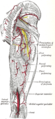
The leg is the entire lower limb of the human body, including the foot, thigh or sometimes even the hip or buttock region. The major bones of the leg are the femur, tibia, and adjacent fibula. The thigh is between the hip and knee, while the calf (rear) and shin (front) are between the knee and foot.

<span class="mw-page-title-main">Femur</span> Thigh bone

The femur, or thigh bone is the only bone in the thigh. The thigh is the region of the lower limb between the hip and the knee. In many four-legged animals the femur is the upper bone of the hindleg.

<span class="mw-page-title-main">Gluteus medius</span> One of the three gluteal muscles

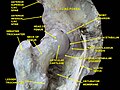
The gluteus medius, one of the three gluteal muscles, is a broad, thick, radiating muscle. It is situated on the outer surface of the pelvis.

<span class="mw-page-title-main">Gluteus minimus</span> Smallest of the three gluteal muscles

The gluteus minimus, or glutæus minimus, the smallest of the three gluteal muscles, is situated immediately beneath the gluteus medius.

<span class="mw-page-title-main">Piriformis muscle</span> One of six small hip muscles in the lateral rotator group

The piriformis muscle is a flat, pyramidally-shaped muscle in the gluteal region of the lower limbs. It is one of the six muscles in the lateral rotator group.

<span class="mw-page-title-main">Obturator internus muscle</span> One of six small hip muscles in the lateral rotator group

The internal obturator muscle or obturator internus muscle originates on the medial surface of the obturator membrane, the ischium near the membrane, and the rim of the pubis.

<span class="mw-page-title-main">Hip</span> Anatomical region between the torso and the legs, holding the buttocks and genital region

In vertebrate anatomy, the hip, or coxa(pl.: coxae) in medical terminology, refers to either an anatomical region or a joint on the outer (lateral) side of the pelvis.

<span class="mw-page-title-main">External obturator muscle</span> One of six small hip muscles in the lateral rotator group

The external obturator muscle or obturator externus muscle is a flat, triangular muscle, which covers the outer surface of the anterior wall of the pelvis.

<span class="mw-page-title-main">Quadratus femoris muscle</span> One of six small hip muscles in the lateral rotator group

The quadratus femoris is a flat, quadrilateral skeletal muscle. Located on the posterior side of the hip joint, it is a strong external rotator and adductor of the thigh, but also acts to stabilize the femoral head in the acetabulum. The quadratus femoris is used in Meyer's muscle pedicle grafting to prevent avascular necrosis of femur head.

<span class="mw-page-title-main">Adductor magnus muscle</span> Muscle in the thigh

The adductor magnus is a large triangular muscle, situated on the medial side of the thigh.

<span class="mw-page-title-main">Gluteal muscles</span> Group of three muscles which make up the buttocks

The gluteal muscles, often called glutes, are a group of three muscles which make up the gluteal region commonly known as the buttocks: the gluteus maximus, gluteus medius and gluteus minimus. The three muscles originate from the ilium and sacrum and insert on the femur. The functions of the muscles include extension, abduction, external rotation, and internal rotation of the hip joint.

<span class="mw-page-title-main">Muscles of the hip</span> Causes movement in the hip

In human anatomy, the muscles of the hip joint are those muscles that cause movement in the hip. Most modern anatomists define 17 of these muscles, although some additional muscles may sometimes be considered. These are often divided into four groups according to their orientation around the hip joint: the gluteal group; the lateral rotator group; the adductor group; and the iliopsoas group.

<span class="mw-page-title-main">Ischium</span> Lower and back region of the hip bone

The ischium forms the lower and back region of the hip bone.

<span class="mw-page-title-main">Lesser trochanter</span> Conical projection from the shaft of the femur

In human anatomy, the lesser trochanter is a conical, posteromedial, bony projection from the shaft of the femur. it serves as the principal insertion site of the iliopsoas muscle.

<span class="mw-page-title-main">Superior gluteal artery</span>

The superior gluteal artery is the terminal branch of the posterior division of the internal iliac artery. It exits the pelvis through the greater sciatic foramen before splitting into a superficial branch and a deep branch.

<span class="mw-page-title-main">Upper extremity of femur</span>

The upper extremity, proximal extremity or superior epiphysis of the femur is the part of the femur closest to the pelvic bone and the trunk. It contains the following structures:

In human anatomy, the body of femur is the almost cylindrical, long part of the femur. It is a little broader above than in the center, broadest and somewhat flattened from before backward below. It is slightly arched, so as to be convex in front, and concave behind, where it is strengthened by a prominent longitudinal ridge, the linea aspera.

<span class="mw-page-title-main">Trochanteric fossa</span>

In mammals including humans, the medial surface of the greater trochanter has at its base a deep depression bounded posteriorly by the intertrochanteric crest, called the trochanteric fossa. This fossa is the point of insertion of four muscles. Moving from the inferior-most to the superior-most, they are: the tendon of the obturator externus muscle, the obturator internus, the superior gemellus and inferior gemellus. The width and depth of the trochanteric fossa varies taxonomically.

<span class="mw-page-title-main">Hip bone</span> Bone of the pelvis

The hip bone is a large flat bone, constricted in the center and expanded above and below. In some vertebrates it is composed of three parts: the ilium, ischium, and the pubis.

<span class="mw-page-title-main">Pelvis</span> Lower torso of the human body

The pelvis is the lower part of the trunk, between the abdomen and the thighs, together with its embedded skeleton.

References

PD-icon.svgThis article incorporates text in the public domain from page 244 of the 20th edition of Gray's Anatomy (1918)

  1. Standring, Susan, editor. Gray’s Anatomy: The Anatomical Basis of Clinical Practice. Forty-First edition, Elsevier Limited, 2016, p. 1327.
  2. Lovell, Wood W.; Winter, Robert B.; Morrissy, Raymond T.; Stuart L. Weinstein (2006). Lovell and Winter's Pediatric Orthopaedics. Lippincott Williams & Wilkins. p. 990. ISBN   978-0-7817-5358-6.