Fifth metatarsal bone

Last updated
Fifth metatarsal bone
Gray288.png
The fifth metatarsal. (Left.)
Ospied-en.svg
Bones of the right foot. Dorsal surface. Fifth metatarsal bone is the yellow bone farthest the right
Details
Identifiers
Latin os metatarsale V
TA2 1502
FMA 24506
Anatomical terms of bone

The fifth metatarsal bone is a long bone in the foot, and is palpable along the distal outer edges of the feet. It is the second smallest of the five metatarsal bones. The fifth metatarsal is analogous to the fifth metacarpal bone in the hand. [1]

Contents

As with the four other metatarsal bones it can be divided into three parts; a base, body and head. The base is the part closest to the ankle and the head is closest to the toes. The narrowed part in the middle is referred to as the body (or shaft) of the bone. The bone is somewhat flat giving it two surfaces; the plantar (towards the sole of the foot) and the dorsal side (the area facing upwards while standing). [1] These surfaces are rough for the attachment of ligaments. The bone is curved longitudinally, so as to be concave below, slightly convex above.

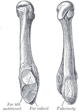
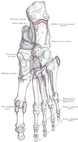
The base articulates behind, by a triangular surface cut obliquely in a transverse direction, with the cuboid; and medially, with the fourth metatarsal. The fifth metatarsal has a rough eminence on the lateral side of its base, known as the tuberosity or the styloid process. The plantar surface of the base is grooved for the tendon of the abductor digiti quinti.

The head articulates with the fifth proximal phalanx, the first bone in the fifth toe.

A strong band of the plantar aponeurosis connects the projecting part of the tuberosity with the lateral process of the tuberosity of the calcaneus.

Proximal fractures

Proximal fractures of 5th metatarsal.jpg

Proximal fractures of the fifth metatarsal are common, [2] and are distinguished by their locations:

Normal anatomy that may simulate a fracture include mainly:

Muscle attachments

Gray268.png
Muscle attachments (seen from above)
Gray269.png
Muscle attachments (seen from below)

The tendon of the fibularis tertius inserts on the medial part of the dorsal surface and the fibularis brevis on the dorsal surface of the tuberosity.

The plantar surface of the base is grooved for the tendon of the abductor digiti quinti, and gives origin to the flexor digiti minimi brevis.

The fourth dorsal interosseus muscle originates from the medial side of shaft. The function of the muscle is to spread the toes. [10]

The third Plantar interosseus muscle originates from the medial side of the base and shaft of the fifth metatarsal. The function of the muscle is to move the fourth toe medially and move the toes together. [10]

The horizontal head of the adductor hallucis from the deep transverse metatarsal ligament, [10] a narrow band which runs across and connects together the heads of all the metatarsal bones.

MuscleDirectionAttachment [11]
Fibularis tertius InsertionDorsal side of the basis
Fibularis brevis InsertionTuberosity
Flexor digiti minimi brevis OriginPlantar surface of the base
Dorsal interossei IV OriginMedial side of the shaft
Plantar interossei III OriginMedial side of the base and shaft
Horizontal head of adductor hallucis OriginDeep transverse metatarsal ligament

Additional images

Related Research Articles

<span class="mw-page-title-main">Foot</span> Anatomical structure found in vertebrates

The foot is an anatomical structure found in many vertebrates. It is the terminal portion of a limb which bears weight and allows locomotion. In many animals with feet, the foot is a separate organ at the terminal part of the leg made up of one or more segments or bones, generally including claws and/or nails.

<span class="mw-page-title-main">Human leg</span> Lower extremity or limb of the human body (foot, lower leg, thigh and hip)

The human leg is the entire lower limb of the human body, including the foot, thigh or sometimes even the hip or buttock region. The major bones of the leg are the femur, tibia, and adjacent fibula. The thigh is between the hip and knee, while the calf (rear) and shin (front) are between the knee and foot.

<span class="mw-page-title-main">Fibularis longus</span> Superficial muscle in the lateral compartment of the leg

In human anatomy, the fibularis longus is a superficial muscle in the lateral compartment of the leg. It acts to tilt the sole of the foot away from the midline of the body (eversion) and to extend the foot downward away from the body at the ankle.

<span class="mw-page-title-main">Cuboid bone</span> Bone of the ankle

In the human body, the cuboid bone is one of the seven tarsal bones of the foot.

<span class="mw-page-title-main">Metatarsal bones</span> Five long bones in the foot

The metatarsal bones, or metatarsus, are a group of five long bones in the midfoot, located between the tarsal bones and the phalanges (toes). Lacking individual names, the metatarsal bones are numbered from the medial side : the first, second, third, fourth, and fifth metatarsal. The metatarsals are analogous to the metacarpal bones of the hand. The lengths of the metatarsal bones in humans are, in descending order, second, third, fourth, fifth, and first. A bovine hind leg has two metatarsals.

<span class="mw-page-title-main">Cuneiform bones</span> Three bones in the human foot

There are three cuneiform ("wedge-shaped") bones in the human foot:

<span class="mw-page-title-main">Calcaneus</span> Bone of the tarsus of the foot

In humans and many other primates, the calcaneus or heel bone is a bone of the tarsus of the foot which constitutes the heel. In some other animals, it is the point of the hock.

<span class="mw-page-title-main">Navicular bone</span> Small bone found in the feet of most mammals

The navicular bone is a small bone found in the feet of most mammals.

<span class="mw-page-title-main">Tarsus (skeleton)</span> Bones of the foot

In the human body, the tarsus is a cluster of seven articulating bones in each foot situated between the lower end of the tibia and the fibula of the lower leg and the metatarsus. It is made up of the midfoot and hindfoot.

<span class="mw-page-title-main">Avulsion fracture</span> Tearing away of a bone by physical trauma

An avulsion fracture is a bone fracture which occurs when a fragment of bone tears away from the main mass of bone as a result of physical trauma. This can occur at the ligament by the application of forces external to the body or at the tendon by a muscular contraction that is stronger than the forces holding the bone together. Generally muscular avulsion is prevented by the neurological limitations placed on muscle contractions. Highly trained athletes can overcome this neurological inhibition of strength and produce a much greater force output capable of breaking or avulsing a bone.

<span class="mw-page-title-main">Dorsal interossei of the foot</span> Four muscles situated between the metatarsal bones

In human anatomy, the dorsal interossei of the foot are four muscles situated between the metatarsal bones.

<span class="mw-page-title-main">Fibularis brevis</span> Shorter and smaller of the fibularis (peroneus) muscles

In human anatomy, the fibularis brevis is a muscle that lies underneath the fibularis longus within the lateral compartment of the leg. It acts to tilt the sole of the foot away from the midline of the body (eversion) and to extend the foot downward away from the body at the ankle.

<span class="mw-page-title-main">Adductor muscles of the hip</span> Group of muscles

The adductor muscles of the hip are a group of muscles in the medial compartment of the thigh mostly used for bringing the thighs together.

<span class="mw-page-title-main">Abductor digiti minimi muscle of foot</span> Muscle which lies along the lateral (outer) border of the foot

The abductor digiti minimi is a muscle which lies along the lateral (outer) border of the foot, and is in relation by its medial margin with the lateral plantar artery, vein and nerves.

<span class="mw-page-title-main">Sole (foot)</span> Bottom part of foot

The sole is the bottom of the foot.

<span class="mw-page-title-main">Fourth metatarsal bone</span>

The fourth metatarsal bone is a long bone in the foot. It is smaller in size than the third metatarsal bone and is the third longest of the five metatarsal bones. The fourth metatarsal is analogous to the fourth metacarpal bone in the hand

<span class="mw-page-title-main">Third metatarsal bone</span>

The third metatarsal bone is a long bone in the foot. It is the second longest metatarsal. The longest being the second metatarsal. The third metatarsal is analogous to the third metacarpal bone in the hand

<span class="mw-page-title-main">Second metatarsal bone</span>

The second metatarsal bone is a long bone in the foot. It is the longest of the metatarsal bones, being prolonged backward and held firmly into the recess formed by the three cuneiform bones. The second metatarsal forms joints with the second proximal phalanx through the metatarsophalangeal joint, the cuneiform bones, third metatarsal and occasionally the first metatarsal bone.

<span class="mw-page-title-main">First metatarsal bone</span>

The first metatarsal bone is the bone in the foot just behind the big toe. The first metatarsal bone is the shortest of the metatarsal bones and by far the thickest and strongest of them.

<span class="mw-page-title-main">Muscles of the thumb</span>

The muscles of the thumb are nine skeletal muscles located in the hand and forearm. The muscles allow for flexion, extension, adduction, abduction and opposition of the thumb. The muscles acting on the thumb can be divided into two groups: The extrinsic hand muscles, with their muscle bellies located in the forearm, and the intrinsic hand muscles, with their muscles bellies located in the hand proper.

References

PD-icon.svgThis article incorporates text in the public domain from page 274 of the 20th edition of Gray's Anatomy (1918)

  1. 1 2 Bojsen-Møller, Finn; Simonsen, Erik B.; Tranum-Jensen, Jørgen (2001). Bevægeapparatets anatomi[Anatomy of the Locomotive Apparatus] (in Danish) (12th ed.). Munksgaard Danmark. p. 246. ISBN   978-87-628-0307-7.
  2. Gary A. Rosenberg; James J. Sferra (September–October 2000). "Treatment Strategies for Acute Fractures and Nonunions of the Proximal Fifth Metatarsal". Journal of the American Academy of Orthopaedic Surgeons. 8 (5): 332–338. doi:10.5435/00124635-200009000-00007. PMID   11029561. S2CID   40453020.
  3. Bica D, Sprouse RA, Armen J (2016). "Diagnosis and Management of Common Foot Fractures". Am Fam Physician. 93 (3): 183–91. PMID   26926612.{{cite journal}}: CS1 maint: multiple names: authors list (link)
  4. "5th Metatarsal". Emergency Care Institute, New South Wales. 2017-09-19. Archived from the original on 2019-07-29. Retrieved 2019-07-29.
  5. "Toe and Forefoot Fractures". OrthoInfo - AAOS. June 2016. Archived from the original on 16 October 2017. Retrieved 15 October 2017.
  6. Robert Silbergleit. "Foot Fracture". Medscape.com. Retrieved 19 October 2011.
  7. Robert Silbergleit. "Foot Fracture". Medscape.com. Retrieved October 19, 2011.
  8. Deniz, G.; Kose, O.; Guneri, B.; Duygun, F. (2014). "Traction apophysitis of the fifth metatarsal base in a child: Iselin's disease". BMJ Case Reports. 2014 (may14 4): bcr2014204687. doi:10.1136/bcr-2014-204687. ISSN   1757-790X. PMC   4025211 . PMID   24832713.
  9. Nwawka, O. Kenechi; Hayashi, Daichi; Diaz, Luis E.; Goud, Ajay R.; Arndt, William F.; Roemer, Frank W.; Malguria, Nagina; Guermazi, Ali (2013). "Sesamoids and accessory ossicles of the foot: anatomical variability and related pathology". Insights into Imaging. 4 (5): 581–593. doi:10.1007/s13244-013-0277-1. ISSN   1869-4101. PMC   3781258 . PMID   24006205.
  10. 1 2 3 Bojsen-Møller, Finn; Simonsen, Erik B.; Tranum-Jensen, Jørgen (2001). Bevægeapparatets anatomi[Anatomy of the Locomotive Apparatus] (in Danish) (12th ed.). Munksgaard Danmark. pp. 300–301. ISBN   978-87-628-0307-7.
  11. Bojsen-Møller, Finn; Simonsen, Erik B.; Tranum-Jensen, Jørgen (2001). Bevægeapparatets anatomi[Anatomy of the Locomotive Apparatus] (in Danish) (12th ed.). Munksgaard Danmark. pp. 364–367. ISBN   978-87-628-0307-7.