| Long bone | |
|---|---|
 Structure of a long bone (femur) | |
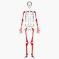
 Long bones in human skeleton (shown in red) | |
| Details | |
| Identifiers | |
| Latin | os longum |
| TA98 | A02.0.00.011 |
| TA2 | 369 |
| FMA | 7474 |
| Anatomical terms of bone | |
The long bones are those that are longer than they are wide. They are one of five types of bones: long, short, flat, irregular and sesamoid. Long bones, especially the femur and tibia, are subjected to most of the load during daily activities and they are crucial for skeletal mobility. They grow primarily by elongation of the diaphysis, with an epiphysis at each end of the growing bone. The ends of epiphyses are covered with hyaline cartilage ("articular cartilage"). The longitudinal growth of long bones is a result of endochondral ossification at the epiphyseal plate. Bone growth in length is stimulated by the production of growth hormone (GH), a secretion of the anterior lobe of the pituitary gland.
Contents
The long bone category includes the femora, tibiae, and fibulae of the legs; the humeri, radii, and ulnae of the arms; metacarpals and metatarsals of the hands and feet, the phalanges of the fingers and toes, and the clavicles or collar bones. The long bones of the human leg make up nearly half of adult height. The other primary skeletal component of height are the vertebrae and skull.
The outside of the bone consists of a layer of connective tissue called the periosteum. Additionally, the outer shell of the long bone is compact bone, then a deeper layer of cancellous bone (spongy bone) which contains in the medullary cavity the bone marrow.