Intramembranous ossification

Last updated

Intramembranous ossification is one of the two essential processes during fetal development of the gnathostome (excluding chondrichthyans such as sharks) skeletal system by which rudimentary bone tissue is created. Intramembranous ossification is also an essential process during the natural healing of bone fractures [1] and the rudimentary formation of bones of the head. [2]

Contents

Transmission electron micrograph of a mesenchymal stem cell that is displaying typical ultrastructural characteristics. MSC high magnification.jpg
Transmission electron micrograph of a mesenchymal stem cell that is displaying typical ultrastructural characteristics.

Unlike endochondral ossification, which is the other process by which bone tissue is created during fetal development, cartilage is not present during intramembranous ossification.

Formation of woven bone

Mesenchymal stem cells within mesenchyme or the medullary cavity of a bone fracture initiate the process of intramembranous ossification. A mesenchymal stem cell, or MSC, is an unspecialized cell that can develop into an osteoblast. Before it begins to develop, the morphological characteristics of a MSC are: A small cell body with a few cell processes that are long and thin; a large, round nucleus with a prominent nucleolus that is surrounded by finely dispersed chromatin particles, giving the nucleus a clear appearance; and a small amount of Golgi apparatus, rough endoplasmic reticulum, mitochondria, and polyribosomes. Furthermore, the mesenchymal stem cells are widely dispersed within an extracellular matrix that is devoid of every type of collagen, except for a few reticular fibrils. [1]

Light micrograph of a nidus consisting of osteoprogenitor cells that are displaying a prominent Golgi apparatus. Bony nidus 1.jpg
Light micrograph of a nidus consisting of osteoprogenitor cells that are displaying a prominent Golgi apparatus.

The process of intramembranous ossification starts when a small group of adjacent MSCs begin to replicate and form a small, dense cluster of cells that is called a nidus. [lower-alpha 1] Once a nidus has been formed the MSCs within it stop replicating. At this point, morphological changes in the MSCs begin to occur: The cell body is now larger and rounder; the long, thin cell processes are no longer present; and the amount of Golgi apparatus and rough endoplasmic reticulum increases. Eventually, all of the cells within the nidus develop into, and display the morphologic characteristics of, an osteoprogenitor cell. [1]

Light micrograph of a nidus consisting of osteoblasts, many are displaying a prominent Golgi apparatus, that have created osteoid at its center. Bony nidus 2.jpg
Light micrograph of a nidus consisting of osteoblasts, many are displaying a prominent Golgi apparatus, that have created osteoid at its center.

At this stage of development, changes in the morphology of the osteoprogenitor cells occur: Their shape becomes more columnar and the amount of Golgi apparatus and rough endoplasmic reticulum increases. Eventually, all of the cells within the nidus develop into, and display the morphologic characteristics of, an osteoblast. Then the osteoblasts create an extracellular matrix containing Type-I collagen fibrils, which is osteoid. The osteoblasts, while lining the periphery of the nidus, continue to form osteoid in the center of the nidus. Some of the osteoblasts become incorporated within the osteoid to become osteocytes. [1]

Light micrograph of an undecalcified nidus consisting of rudimentary bone tissue that is lined by numerous osteoblasts. Bony nidus 3.jpg
Light micrograph of an undecalcified nidus consisting of rudimentary bone tissue that is lined by numerous osteoblasts.

At this point, the osteoid becomes mineralized resulting in a nidus consisting of mineralized osteoid that contains osteocytes and is lined by active osteoblasts. The nidus, that began as a diffuse collection of MSCs, has developed into woven bone, the most rudimentary bone tissue. [1]

Formation of lamellar bone

The first step in the process is the formation of bone spicules which eventually fuse with each other and become trabeculae. The periosteum is formed and bone growth continues at the surface of trabeculae. Much like spicules, the increasing growth of trabeculae result in interconnection and this network is called woven bone. Eventually, woven bone is replaced by lamellar bone .

Formation of bone spicules

Embryologic mesenchymal cells (MSC) condense into layers of vascularized primitive connective tissue. Certain mesenchymal cells group together, usually near or around blood vessels, and differentiate into osteogenic cells which deposit bone matrix constitutively. These aggregates of bony matrix are called bone spicules. Separate mesenchymal cells differentiate into osteoblasts, which line up along the surface of the spicule and secrete more osteoid, which increases the size of the spicule.

Formation of trabecular bone

As the spicules continue to grow, they fuse with adjacent spicules and this results in the formation of trabeculae . When osteoblasts become trapped in the matrix they secrete, they differentiate into osteocytes. Osteoblasts continue to line up on the surface which increases the size. As growth continues, trabeculae become interconnected and trabecular bone is formed. The term primary spongiosa is also used to refer to the initial trabecular network.

Primary centre of ossification

The periosteum is formed around the trabeculae by differentiating mesenchymal cells. The primary center of ossification is the area where bone growth occurs between the periosteum and the bone. Osteogenic cells that originate from the periosteum increase appositional growth and a bone collar is formed. The bone collar is eventually mineralized and lamellar bone is formed.

Formation of osteon

Osteons are components or principal structures of compact bone. During the formation of bone spicules, cytoplasmic processes from osteoblasts interconnect. This becomes the canaliculi of osteons. Since bone spicules tend to form around blood vessels, the perivascular space is greatly reduced as the bone continues to grow. When replacement to compact bone occurs, this blood vessel becomes the central canal of the osteon.

Examples in the human body

The following bones develop in humans via Intramembranous ossification: [3]

Other bone that formed by intramembranous ossification are: cortices of tubular and flat bones as well as the calvaria, upper facial bones, tympanic temporal bone, vomer, and medial pterygoid process. [4]

See also

Footnotes

  1. Nidus is Latin for "nest". In tissue, a nidus resembles a nest morphologically, with respect to its appearance, and functionally, because it is a site where cell development occurs.

Related Research Articles

<span class="mw-page-title-main">Bone</span> Rigid organs that constitute part of the endoskeleton of vertebrates

A bone is a rigid organ that constitutes part of the skeleton in most vertebrate animals. Bones protect the various other organs of the body, produce red and white blood cells, store minerals, provide structure and support for the body, and enable mobility. Bones come in a variety of shapes and sizes and have complex internal and external structures. They are lightweight yet strong and hard and serve multiple functions.

<span class="mw-page-title-main">Bone healing</span> Healing from bone injury

Bone healing, or fracture healing, is a proliferative physiological process in which the body facilitates the repair of a bone fracture.

<span class="mw-page-title-main">Osteoblast</span> Cells secreting extracellular matrix

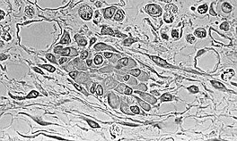
Osteoblasts are cells with a single nucleus that synthesize bone. However, in the process of bone formation, osteoblasts function in groups of connected cells. Individual cells cannot make bone. A group of organized osteoblasts together with the bone made by a unit of cells is usually called the osteon.

<span class="mw-page-title-main">Osteoid</span> Unmineralized, organic portion of the bone matrix

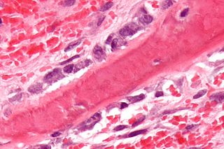
In histology, osteoid is the unmineralized, organic portion of the bone matrix that forms prior to the maturation of bone tissue. Osteoblasts begin the process of forming bone tissue by secreting the osteoid as several specific proteins. When the osteoid becomes mineralized, it and the adjacent bone cells have developed into new bone tissue.

<span class="mw-page-title-main">Osteoclast</span> Cell that breaks down bone tissue

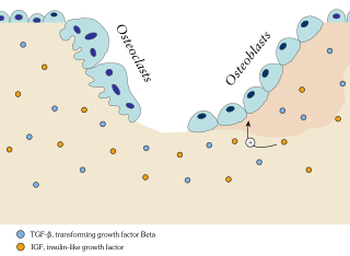
An osteoclast is a type of bone cell that breaks down bone tissue. This function is critical in the maintenance, repair, and remodeling of bones of the vertebral skeleton. The osteoclast disassembles and digests the composite of hydrated protein and mineral at a molecular level by secreting acid and a collagenase, a process known as bone resorption. This process also helps regulate the level of blood calcium.

<span class="mw-page-title-main">Periosteum</span> Membrane covering outer surface of bones

The periosteum is a membrane that covers the outer surface of all bones, except at the articular surfaces of long bones. Endosteum lines the inner surface of the medullary cavity of all long bones.

<span class="mw-page-title-main">Osteocyte</span> Mature osteoblasts which helps in communication between cells and also in molecular synthesis

An osteocyte, an oblate shaped type of bone cell with dendritic processes, is the most commonly found cell in mature bone. It can live as long as the organism itself. The adult human body has about 42 billion of them. Osteocytes do not divide and have an average half life of 25 years. They are derived from osteoprogenitor cells, some of which differentiate into active osteoblasts. Osteoblasts/osteocytes develop in mesenchyme.

<span class="mw-page-title-main">Chondrocyte</span> Cell that makes up cartilage

Chondrocytes are the only cells found in healthy cartilage. They produce and maintain the cartilaginous matrix, which consists mainly of collagen and proteoglycans. Although the word chondroblast is commonly used to describe an immature chondrocyte, the term is imprecise, since the progenitor of chondrocytes can differentiate into various cell types, including osteoblasts.

<span class="mw-page-title-main">Endochondral ossification</span> Cartilaginous bone development that forms the long bones

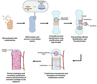
Endochondral ossification is one of the two essential processes during fetal development of the mammalian skeletal system by which bone tissue is produced. Unlike intramembranous ossification, the other process by which bone tissue is produced, cartilage is present during endochondral ossification. Endochondral ossification is also an essential process during the rudimentary formation of long bones, the growth of the length of long bones, and the natural healing of bone fractures.

<span class="mw-page-title-main">Osteon</span> Fundamental anatomical unit of compact bone

In osteology, the osteon or haversian system is the fundamental functional unit of much compact bone. Osteons are roughly cylindrical structures that are typically between 0.25 mm and 0.35 mm in diameter. Their length is often hard to define, but estimates vary from several millimeters to around 1 centimeter. They are present in many bones of most mammals and some bird, reptile, and amphibian species.

<span class="mw-page-title-main">Ossification</span> Development process in bones

Ossification in bone remodeling is the process of laying down new bone material by cells named osteoblasts. It is synonymous with bone tissue formation. There are two processes resulting in the formation of normal, healthy bone tissue: Intramembranous ossification is the direct laying down of bone into the primitive connective tissue (mesenchyme), while endochondral ossification involves cartilage as a precursor.

<span class="mw-page-title-main">Bone resorption</span> Medical condition

Bone resorption is resorption of bone tissue, that is, the process by which osteoclasts break down the tissue in bones and release the minerals, resulting in a transfer of calcium from bone tissue to the blood.

<span class="mw-page-title-main">Chondroblast</span> Mesenchymal progenitor cell that forms a chondrocyte

Chondroblasts, or perichondrial cells, is the name given to mesenchymal progenitor cells in situ which, from endochondral ossification, will form chondrocytes in the growing cartilage matrix. Another name for them is subchondral cortico-spongious progenitors. They have euchromatic nuclei and stain by basic dyes.

<span class="mw-page-title-main">Osteonecrosis of the jaw</span> Medical condition

Osteonecrosis of the jaw (ONJ) is a severe bone disease (osteonecrosis) that affects the jaws. Various forms of ONJ have been described since 1861, and a number of causes have been suggested in the literature.

<span class="mw-page-title-main">Bone remodeling</span> Continuous turnover of bone matrix and mineral

In osteology, bone remodeling or bone metabolism is a lifelong process where mature bone tissue is removed from the skeleton and new bone tissue is formed. These processes also control the reshaping or replacement of bone following injuries like fractures but also micro-damage, which occurs during normal activity. Remodeling responds also to functional demands of the mechanical loading.

<span class="mw-page-title-main">Sp7 transcription factor</span> Protein-coding gene in the species Homo sapiens

Transcription factor Sp7, also called osterix (Osx), is a protein that in humans is encoded by the SP7 gene. It is a member of the Sp family of zinc-finger transcription factors It is highly conserved among bone-forming vertebrate species It plays a major role, along with Runx2 and Dlx5 in driving the differentiation of mesenchymal precursor cells into osteoblasts and eventually osteocytes. Sp7 also plays a regulatory role by inhibiting chondrocyte differentiation maintaining the balance between differentiation of mesenchymal precursor cells into ossified bone or cartilage. Mutations of this gene have been associated with multiple dysfunctional bone phenotypes in vertebrates. During development, a mouse embryo model with Sp7 expression knocked out had no formation of bone tissue. Through the use of GWAS studies, the Sp7 locus in humans has been strongly associated with bone mass density. In addition there is significant genetic evidence for its role in diseases such as Osteogenesis imperfecta (OI).

<span class="mw-page-title-main">Osteochondroprogenitor cell</span>

Osteochondroprogenitor cells are progenitor cells that arise from mesenchymal stem cells (MSC) in the bone marrow. They have the ability to differentiate into osteoblasts or chondrocytes depending on the signalling molecules they are exposed to, giving rise to either bone or cartilage respectively. Osteochondroprogenitor cells are important for bone formation and maintenance.

Wolff's law, developed by the German anatomist and surgeon Julius Wolff (1836–1902) in the 19th century, states that bone in a healthy animal will adapt to the loads under which it is placed. If loading on a particular bone increases, the bone will remodel itself over time to become stronger to resist that sort of loading. The internal architecture of the trabeculae undergoes adaptive changes, followed by secondary changes to the external cortical portion of the bone, perhaps becoming thicker as a result. The inverse is true as well: if the loading on a bone decreases, the bone will become less dense and weaker due to the lack of the stimulus required for continued remodeling. This reduction in bone density (osteopenia) is known as stress shielding and can occur as a result of a hip replacement. The normal stress on a bone is shielded from that bone by being placed on a prosthetic implant.

<span class="mw-page-title-main">Mesenchymal stem cell</span> Multipotent, non-hematopoietic adult stem cells present in multiple tissues

Mesenchymal stem cells (MSCs) also known as mesenchymal stromal cells or medicinal signaling cells are multipotent stromal cells that can differentiate into a variety of cell types, including osteoblasts, chondrocytes, myocytes and adipocytes.

Craniofacial regeneration refers to the biological process by which the skull and face regrow to heal an injury. This page covers birth defects and injuries related to the craniofacial region, the mechanisms behind the regeneration, the medical application of these processes, and the scientific research conducted on this specific regeneration. This regeneration is not to be confused with tooth regeneration. Craniofacial regrowth is broadly related to the mechanisms of general bone healing.

References

  1. 1 2 3 4 5 Brighton, Carl T.; Robert M. Hunt (1991). "Early histological and ultrastructural changes in medullary fracture callus". Journal of Bone and Joint Surgery . 73-A (6): 832–847.
  2. Netter, Frank H. (1987). Musculoskeletal system: anatomy, physiology, and metabolic disorders. Summit, New Jersey: Ciba-Geigy Corporation. p. 129. ISBN   0-914168-88-6.
  3. "6.4 Bone Formation and Development – Anatomy and Physiology". opentextbc.ca. Retrieved 5 May 2018.
  4. Ihde, Lauren L.; Forrester, Deborah M.; Gottsegen, Christopher J.; Masih, Sulabha; Patel, Dakshesh B.; Vachon, Linda A.; White, Eric A.; Matcuk, George R. (November 2011). "Sclerosing Bone Dysplasias: Review and Differentiation from Other Causes of Osteosclerosis". RadioGraphics. 31 (7): 1865–1882. doi:10.1148/rg.317115093. ISSN   0271-5333.