Ultrastructure

Last updated
The ultrastructure of a single bacterial cell (Bacillus subtilis). The scale bar is 200 nm. Bacillus subtilis.jpg
The ultrastructure of a single bacterial cell ( Bacillus subtilis ). The scale bar is 200 nm.

Ultrastructure (or ultra-structure) is the architecture of cells and biomaterials that is visible at higher magnifications than found on a standard optical light microscope. This traditionally meant the resolution and magnification range of a conventional transmission electron microscope (TEM) when viewing biological specimens such as cells, tissue, or organs. Ultrastructure can also be viewed with scanning electron microscopy and super-resolution microscopy, although TEM is a standard histology technique for viewing ultrastructure. Such cellular structures as organelles, which allow the cell to function properly within its specified environment, can be examined at the ultrastructural level.

Contents

Ultrastructure, along with molecular phylogeny, is a reliable phylogenetic way of classifying organisms. [1] Features of ultrastructure are used industrially to control material properties and promote biocompatibility.

History

In 1931, German engineers Max Knoll and Ernst Ruska invented the first electron microscope. [2] With the development and invention of this microscope, the range of observable structures that were able to be explored and analyzed increased immensely, as biologists became progressively interested in the submicroscopic organization of cells. This new area of research concerned itself with substructure, also known as the ultrastructure. [3]

Applications

Many scientists use ultrastructural observations to study the following, including but not limited to:

Biology

A common ultrastructural feature found in plant cells is the formation of calcium oxalate crystals. [9] It has been theorized that these crystals function to store calcium within the cell until it is needed for growth or development. [10]

Calcium oxalate crystals can also form in animals, and kidney stones are a form of these ultrastructural features. Theoretically, nanobacteria could be used to decrease the formation of calcium oxalate kidney stones. [11]

Engineering

Controlling ultrastructure has engineering uses for controlling the behavior of cells. Cells respond readily to changes in their extracellular matrix (ECM), so manufacturing materials to mimic ECM allows for increased control over the cell cycle and protein expression. [12]

Many cells, such as plants, produce calcium oxalate crystals, and these crystals are usually considered ultrastructural components of plant cells. Calcium oxalate is a material that is used to manufacture ceramic glazes [6], and it also has biomaterial properties. For culturing cells and tissue engineering, this crystal is found in fetal bovine serum, and is an important aspect of the extracellular matrix for culturing cells. [13]  

Ultrastructure is an important factor to consider when engineering dental implants. Since these devices interface directly with bone, their incorporation to surrounding tissue is necessary to optimal device function. It has been found that applying a load to a healing dental implant allows for increased osseointegration with facial bones. [14] Analyzing the ultrastructure surrounding an implant is useful in determining how biocompatible it is and how the body reacts to it. One study found implanting granules of a biomaterial derived from pig bone caused the human body to incorporate the material into its ultrastructure and form new bone. [15]

Hydroxyapatite is a biomaterial used to interface medical devices directly to bone by ultrastructure. Grafts can be created along with 𝛃-tricalcium phosphate, and it has been observed that surrounding bone tissue with incorporate the new material into its extracellular matrix. [16] Hydroxyapatite is a highly biocompatible material, and its ultrastructural features, such as crystalline orientation, can be controlled carefully to ensure optimal biocompatibility. [17] Proper crystal fiber orientation can make introduced minerals, like hydroxyapatite, more similar to the biological materials they intend to replace. Controlling ultrastructural features makes obtaining specific material properties possible.

Related Research Articles

<span class="mw-page-title-main">Osteoblast</span> Cells secreting extracellular matrix

Osteoblasts are cells with a single nucleus that synthesize bone. However, in the process of bone formation, osteoblasts function in groups of connected cells. Individual cells cannot make bone. A group of organized osteoblasts together with the bone made by a unit of cells is usually called the osteon.

<span class="mw-page-title-main">Hydroxyapatite</span> Naturally occurring mineral form of calcium apatite

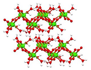
Hydroxyapatite is a naturally occurring mineral form of calcium apatite with the formula Ca5(PO4)3(OH), often written Ca10(PO4)6(OH)2 to denote that the crystal unit cell comprises two entities. It is the hydroxyl endmember of the complex apatite group. The OH ion can be replaced by fluoride or chloride, producing fluorapatite or chlorapatite. It crystallizes in the hexagonal crystal system. Pure hydroxyapatite powder is white. Naturally occurring apatites can, however, also have brown, yellow, or green colorations, comparable to the discolorations of dental fluorosis.

<span class="mw-page-title-main">Bioglass 45S5</span>

Bioglass 45S5 or calcium sodium phosphosilicate, is a bioactive glass specifically composed of 45 wt% SiO2, 24.5 wt% CaO, 24.5 wt% Na2O, and 6.0 wt% P2O5. Typical applications of Bioglass 45S5 include: bone grafting biomaterials, repair of periodontal defects, cranial and maxillofacial repair, wound care, blood loss control, stimulation of vascular regeneration, and nerve repair.

<span class="mw-page-title-main">Bioactive glass</span>

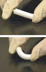
Bioactive glasses are a group of surface reactive glass-ceramic biomaterials and include the original bioactive glass, Bioglass. The biocompatibility and bioactivity of these glasses has led them to be used as implant devices in the human body to repair and replace diseased or damaged bones. Most bioactive glasses are silicate-based glasses that are degradable in body fluids and can act as a vehicle for delivering ions beneficial for healing. Bioactive glass is differentiated from other synthetic bone grafting biomaterials, in that it is the only one with anti-infective and angiogenic properties.

<span class="mw-page-title-main">Bone grafting</span> Bone transplant

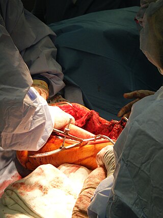
Bone grafting is a surgical procedure that replaces missing bone in order to repair bone fractures that are extremely complex, pose a significant health risk to the patient, or fail to heal properly. Some small or acute fractures can be cured without bone grafting, but the risk is greater for large fractures like compound fractures.

<span class="mw-page-title-main">Tricalcium phosphate</span> Chemical compound

Tricalcium phosphate (sometimes abbreviated TCP), more commonly known as Calcium phosphate, is a calcium salt of phosphoric acid with the chemical formula Ca3(PO4)2. It is also known as tribasic calcium phosphate and bone phosphate of lime (BPL). It is a white solid of low solubility. Most commercial samples of "tricalcium phosphate" are in fact hydroxyapatite.

<span class="mw-page-title-main">Dicalcium phosphate</span> Chemical compound

Dicalcium phosphate is the calcium phosphate with the formula CaHPO4 and its dihydrate. The "di" prefix in the common name arises because the formation of the HPO42– anion involves the removal of two protons from phosphoric acid, H3PO4. It is also known as dibasic calcium phosphate or calcium monohydrogen phosphate. Dicalcium phosphate is used as a food additive, it is found in some toothpastes as a polishing agent and is a biomaterial.

<span class="mw-page-title-main">Biomaterial</span> Any substance that has been engineered to interact with biological systems for a medical purpose

A biomaterial is a substance that has been engineered to interact with biological systems for a medical purpose, either a therapeutic or a diagnostic one. The corresponding field of study, called biomaterials science or biomaterials engineering, is about fifty years old. It has experienced steady and strong growth over its history, with many companies investing large amounts of money into the development of new products. Biomaterials science encompasses elements of medicine, biology, chemistry, tissue engineering and materials science.

<span class="mw-page-title-main">Foreign body reaction</span> Medical condition

A foreign body reaction (FBR) is a typical tissue response to a foreign body within biological tissue. It usually includes the formation of a foreign body granuloma. Tissue-encapsulation of an implant is an example, as is inflammation around a splinter. Foreign body granuloma formation consists of protein adsorption, macrophages, multinucleated foreign body giant cells, fibroblasts, and angiogenesis. It has also been proposed that the mechanical property of the interface between an implant and its surrounding tissues is critical for the host response.

Biomimetic materials are materials developed using inspiration from nature. This may be useful in the design of composite materials. Natural structures have inspired and innovated human creations. Notable examples of these natural structures include: honeycomb structure of the beehive, strength of spider silks, bird flight mechanics, and shark skin water repellency. The etymological roots of the neologism "biomimetic" derive from Greek, since bios means "life" and mimetikos means "imitative".

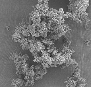
Amorphous calcium phosphate (ACP) is a glassy solid that is formed from the chemical decomposition of a mixture of dissolved phosphate and calcium salts (e.g. (NH4)2HPO4 + Ca(NO3)2). The resulting amorphous mixture consists mostly of calcium and phosphate, but also contains varying amounts of water and hydrogen and hydroxide ions, depending on the synthesis conditions. Such mixtures are also known as calcium phosphate cement.

<span class="mw-page-title-main">Clemens van Blitterswijk</span>

Clemens A. van Blitterswijk is a Dutch tissue engineer who contributed to the use of biomaterials to heal bone injuries, especially using osteoinductive ceramics. In collaboration with Jan de Boer and others, he has contributed to screening microtextures to study cell-biomaterial interactions, an approach termed materiomics.

<span class="mw-page-title-main">Bioceramic</span> Type of ceramic materials that are biocompatible

Bioceramics and bioglasses are ceramic materials that are biocompatible. Bioceramics are an important subset of biomaterials. Bioceramics range in biocompatibility from the ceramic oxides, which are inert in the body, to the other extreme of resorbable materials, which are eventually replaced by the body after they have assisted repair. Bioceramics are used in many types of medical procedures. Bioceramics are typically used as rigid materials in surgical implants, though some bioceramics are flexible. The ceramic materials used are not the same as porcelain type ceramic materials. Rather, bioceramics are closely related to either the body's own materials or are extremely durable metal oxides.

<span class="mw-page-title-main">Artificial bone</span> Bone-like material

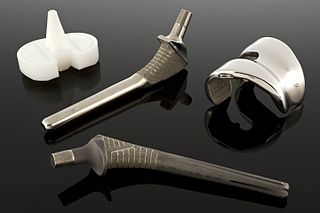
Artificial bone refers to bone-like material created in a laboratory that can be used in bone grafts, to replace human bone that was lost due to severe fractures, disease, etc.

Socket preservation or alveolar ridge preservation is a procedure to reduce bone loss after tooth extraction. After tooth extraction, the jaw bone has a natural tendency to become narrow, and lose its original shape because the bone quickly resorbs, resulting in 30–60% loss in bone volume in the first six months. Bone loss, can compromise the ability to place a dental implant, or its aesthetics and functional ability.

<span class="mw-page-title-main">Mineralized tissues</span> Biological tissues incorporating minerals

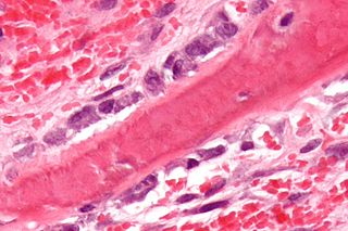
Mineralized tissues are biological tissues that incorporate minerals into soft matrices. Typically these tissues form a protective shield or structural support. Bone, mollusc shells, deep sea sponge Euplectella species, radiolarians, diatoms, antler bone, tendon, cartilage, tooth enamel and dentin are some examples of mineralized tissues.

Octacalcium phosphate (sometimes referred to as OCP) is a form of calcium phosphate with formula Ca8H2(PO4)6·5H2O. OCP may be a precursor to tooth enamel, dentine, and bones. OCP is a precursor of hydroxyapatite (HA), an inorganic biomineral that is important in bone growth. OCP has garnered lots of attention due to its inherent biocompatibility. While OCP exhibits good properties in terms of bone growth, very stringent synthesis requirements make it difficult for mass productions, but nevertheless has shown promise not only in-vitro, but also in in-vivo clinical case studies.

<span class="mw-page-title-main">Surface modification of biomaterials with proteins</span>

Biomaterials are materials that are used in contact with biological systems. Biocompatibility and applicability of surface modification with current uses of metallic, polymeric and ceramic biomaterials allow alteration of properties to enhance performance in a biological environment while retaining bulk properties of the desired device.

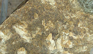
Tetracalcium phosphate is the compound Ca4(PO4)2O, (4CaO·P2O5). It is the most basic of the calcium phosphates, and has a Ca/P ratio of 2, making it the most phosphorus poor phosphate. It is found as the mineral hilgenstockite, which is formed in industrial phosphate rich slag (called "Thomas slag"). This slag was used as a fertiliser due to the higher solubility of tetracalcium phosphate relative to apatite minerals. Tetracalcium phosphate is a component in some calcium phosphate cements that have medical applications.

Crystallopathy is a harmful state or disease associated with the formation and aggregation of crystals in tissues or cavities, or in other words, a heterogeneous group of diseases caused by intrinsic or environmental microparticles or crystals, promoting tissue inflammation and scarring.

References

  1. Laura Wegener Parfrey; Erika Barbero; Elyse Lasser; Micah Dunthorn; Debashish Bhattacharya; David J Patterson; Laura A Katz (December 2006). "Evaluating support for the current classification of eukaryotic diversity". PLOS Genetics . 2 (12): e220. doi: 10.1371/JOURNAL.PGEN.0020220 . ISSN   1553-7390. PMC   1713255 . PMID   17194223. Wikidata   Q21090155.
  2. Masters, Barry R (March 2009). "History of the Electron Microscope in Cell Biology" (PDF). Encyclopedia of Life Sciences (ELS). Chichester: John Wiley & Sons, Ltd. doi:10.1002/9780470015902.a0021539. ISBN   9780470016176.
  3. Brieger, E.M. (1963). "Ultrastructure of the Cell". Structure and Ultrastructure of Microorganisms. Elsevier. p. 1–7. doi:10.1016/b978-0-12-134350-7.50005-8. ISBN   978-0-12-134350-7.
  4. Eyden, B.; Banerjee, S.S.; Ru, Y.; Liberski, P. (2014). The Ultrastructure of Human Tumours: Applications in Diagnosis and Research. Springer Berlin Heidelberg. ISBN   978-3-642-39168-2.
  5. Musser, Robert L.; Thomas, Shirley A.; Wise, Robert R.; Peeler, Thomas C.; Naylor, Aubrey W. (1984-04-01). "Chloroplast Ultrastructure, Chlorophyll Fluorescence, and Pigment Composition in Chilling-Stressed Soybeans". Plant Physiology. 74 (4): 749–754. doi:10.1104/pp.74.4.749. ISSN   0032-0889. PMC   1066762 . PMID   16663504.
  6. Moreira, Carolina A.; Dempster, David W.; Baron, Roland (2000). "Anatomy and Ultrastructure of Bone – Histogenesis, Growth and Remodeling". Endotext. South Dartmouth (MA): MDText.com, Inc. PMID   25905372.
  7. Cramer, Elisabeth M.; Norol, Françoise; Guichard, Josette; Breton-Gorius, Janine; Vainchenker, William; Massé, Jean-Marc; Debili, Najet (1997-04-01). "Ultrastructure of Platelet Formation by Human Megakaryocytes Cultured With the Mpl Ligand". Blood. 89 (7): 2336–2346. doi: 10.1182/blood.V89.7.2336 . ISSN   1528-0020. PMID   9116277. S2CID   7757033.
  8. Ferreira, Adelina; Dolder, Heidi (1990-01-06). "Sperm ultrastructure and spermatogenesis in the lizard, Tropidurus itambere" (PDF). Biocell. 27 (3): 353–362. ISSN   0327-9545. PMID   15002752.
  9. Prychid, C. J.; Jabaily, R. S.; Rudall, P. J. (2008-03-13). "Cellular Ultrastructure and Crystal Development in Amorphophallus (Araceae)". Annals of Botany. 101 (7): 983–995. doi:10.1093/aob/mcn022. ISSN   0305-7364. PMC   2710233 . PMID   18285357.
  10. Tilton, V. R.; Horner, H. T. (1980). "Calcium Oxalate Raphide Crystals and Crystalliferous Idioblasts in the Carpels of Ornithogalum caudatum". Annals of Botany. 46 (5): 533–539. doi:10.1093/oxfordjournals.aob.a085951. ISSN   1095-8290.
  11. Goldfarb, David S. (2004-10-19). "Microorganisms and Calcium Oxalate Stone Disease". Nephron Physiology. 98 (2): 48–54. doi:10.1159/000080264. ISSN   1660-2137. PMID   15499215. S2CID   29369994.
  12. Khademhosseini, Ali (2008). Micro and nanoengineering of the cell microenvironment: technologies and applications. Boston: Artech House. ISBN   978-1-59693-149-7.
  13. Pedraza, Claudio E.; Chien, Yung‐Ching; McKee, Marc D. (2008). "Calcium oxalate crystals in fetal bovine serum: Implications for cell culture, phagocytosis and biomineralization studies in vitro". Journal of Cellular Biochemistry. 103 (5): 1379–1393. doi:10.1002/jcb.21515. ISSN   0730-2312. PMID   17879965. S2CID   43217705.
  14. Meyer, U.; Joos, U.; Mythili, J.; Stamm, T.; Hohoff, A.; Fillies, T.; Stratmann, U.; Wiesmann, H.P. (2004). "Ultrastructural characterization of the implant/bone interface of immediately loaded dental implants". Biomaterials. 25 (10): 1959–1967. doi:10.1016/j.biomaterials.2003.08.070. PMID   14738860.
  15. Orsini, Giovanna; Scarano, Antonio; Piattelli, Maurizio; Piccirilli, Marcello; Caputi, Sergio; Piattelli, Adriano (2006). "Histologic and Ultrastructural Analysis of Regenerated Bone in Maxillary Sinus Augmentation Using a Porcine Bone–Derived Biomaterial". Journal of Periodontology. 77 (12): 1984–1990. doi:10.1902/jop.2006.060181. ISSN   0022-3492. PMID   17209782.
  16. Fujita, Rumi; Yokoyama, Atsuro; Nodasaka, Yoshinobu; Kohgo, Takao; Kawasaki, Takao (2003). "Ultrastructure of ceramic-bone interface using hydroxyapatite and β-tricalcium phosphate ceramics and replacement mechanism of β-tricalcium phosphate in bone". Tissue and Cell. 35 (6): 427–440. doi:10.1016/S0040-8166(03)00067-3. PMID   14580356.
  17. Zhuang, Zhi; Miki, Takuya; Yumoto, Midori; Konishi, Toshiisa; Aizawa, Mamoru (2012). "Ultrastructural Observation of Hydroxyapatite Ceramics with Preferred Orientation to a-plane Using High-resolution Transmission Electron Microscopy". Procedia Engineering. 36: 121–127. doi: 10.1016/j.proeng.2012.03.019 .