Calvaria (skull)

Last updated
Calvaria (skull)
Kort-lang-skalle.gif
Details
Part ofThe calvaria is the top part of the skull
Identifiers
Latin calvaria
TA98 A02.1.00.032
TA2 436
FMA 52800
Anatomical terms of bone

The calvaria is the top part of the skull. It is the superior part of the neurocranium and covers the cranial cavity containing the brain. It forms the main component of the skull roof.

Contents

The calvaria is made up of the superior portions of the frontal bone, occipital bone, and parietal bones. [1] In the human skull, the sutures between the bones normally remain flexible during the first few years of postnatal development, and fontanelles are palpable. Premature complete ossification of these sutures is called craniosynostosis.

In Latin, the word calvaria is used as a feminine noun with plural calvariae; however, many medical texts incorrectly list the word as calvarium, a neuter Latin noun with plural calvaria.[ citation needed ]

Structure

Resistance structures of calvaria Functional architecture of cranium.jpg
Resistance structures of calvaria

The outer surface of the skull possesses a number of landmarks. The point at which the frontal bone and the two parietal bones meet is known as the bregma. The point at which the two parietal bones and the occipital bone meet is known as the lambda. Not only do these landmarks indicate the fontanelle in newborns, they also act as reference points in medicine and surgery.

The inner surface of the skull-cap is concave and presents depressions for the convolutions of the cerebrum, together with numerous furrows for the lodgement of branches of the meningeal vessels. Along the middle line is a longitudinal groove, narrow in front, where it commences at the frontal crest, but broader behind; it lodges the superior sagittal sinus, and its margins afford attachment to the falx cerebri. On either side of it are several depressions for the arachnoid granulations, and at its back part, the openings of the parietal foramina when these are present.

It is crossed in front by the coronal suture and behind by the lambdoid suture, while the sagittal suture lies in the medial plane between the parietal bones.

Layers

The outer layer of the skull has been removed and shows the diploic veins and inner layer. Gray564.png
The outer layer of the skull has been removed and shows the diploic veins and inner layer.

Most bones of the calvaria consist of internal and external tables or layers of compact bone, separated by diploë. The diploë is cancellous bone containing red bone marrow during life, through which run canals formed by diploic veins. The diploë in a dried calvaria is not red because the protein was removed during preparation of the cranium. The internal table of bone is thinner than the external table, and in some areas there is only a thin plate of compact bone with no diploë. [2] Calvarial bones are supplied by endosteal and periosteal sheaths which are innervated by the nociceptors, sensory, sympathetic, and parasympathetic nerves. Horizontal section of the mouse pups showed that the density of nerve fibers was highest in the region of forehead, temples, and the back of head which crossing the frontal, parietal, and interparietal bones. In the calvarial innervation in the adult mouse, CGRP-labeled fibers and peripherin were seen in the sutures, emissary canals, and bone marrow but not in diploe. Nerve fibers passing through the emissary canals and cavity of bone marrow provided the branches of periosteal and dural nerves whereas fibers from the sutures gave out to the dural nerves. [3]

Development

In the fetus, the formation of the calvaria involves a process known as intramembranous ossification.

In many translations of the Gospels, Jesus is killed in a place named "Calvary", a reference to this body part.

Related Research Articles

<span class="mw-page-title-main">Skull</span> Bony structure that forms the head in vertebrates

The skull is a bone protective cavity for the brain. The skull is composed of four types of bone i.e., cranial bones, facial bones, ear ossicles and hyoid bone. However two parts are more prominent: the cranium and the mandible. In humans, these two parts are the neurocranium and the viscerocranium that includes the mandible as its largest bone. The skull forms the anterior-most portion of the skeleton and is a product of cephalisation—housing the brain, and several sensory structures such as the eyes, ears, nose, and mouth. In humans these sensory structures are part of the facial skeleton.

<span class="mw-page-title-main">Fontanelle</span> Anatomical feature of the infant human skull

A fontanelle is an anatomical feature of the infant human skull comprising soft membranous gaps (sutures) between the cranial bones that make up the calvaria of a fetus or an infant. Fontanelles allow for stretching and deformation of the neurocranium both during birth and later as the brain expands faster than the surrounding bone can grow. Premature complete ossification of the sutures is called craniosynostosis.

<span class="mw-page-title-main">Facial nerve</span> Cranial nerve VII, for the face and tasting

The facial nerve, also known as the seventh cranial nerve, cranial nerve VII, or simply CN VII, is a cranial nerve that emerges from the pons of the brainstem, controls the muscles of facial expression, and functions in the conveyance of taste sensations from the anterior two-thirds of the tongue. The nerve typically travels from the pons through the facial canal in the temporal bone and exits the skull at the stylomastoid foramen. It arises from the brainstem from an area posterior to the cranial nerve VI and anterior to cranial nerve VIII.

Articles related to anatomy include:

<span class="mw-page-title-main">Occipital bone</span> Saucer-shaped membrane bone situated at the back and lower part of the cranium

The occipital bone is a cranial dermal bone and the main bone of the occiput. It is trapezoidal in shape and curved on itself like a shallow dish. The occipital bone overlies the occipital lobes of the cerebrum. At the base of the skull in the occipital bone, there is a large oval opening called the foramen magnum, which allows the passage of the spinal cord.

<span class="mw-page-title-main">Parietal bone</span> Bone in the human skull which, when joined together, forms the sides and roof of the cranium

The parietal bones are two bones in the skull which, when joined at a fibrous joint, form the sides and roof of the cranium. In humans, each bone is roughly quadrilateral in form, and has two surfaces, four borders, and four angles. It is named from the Latin paries (-ietis), wall.

<span class="mw-page-title-main">Scalp</span> Anatomical area bordered by the human face at the front, and by the neck at the sides and back

The scalp is the anatomical area bordered by the human face at the front, and by the neck at the sides and back.

<span class="mw-page-title-main">Diploic veins</span> Large valveless veins in the skull

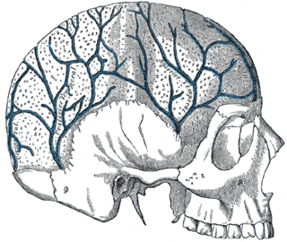
The diploic veins are large, thin-walled valveless veins that channel in the diploë between the inner and outer layers of the cortical bone in the skull. They are lined by a single layer of endothelium supported by elastic tissue. They develop fully by the age of two years. The diploic veins drain this area into the dural venous sinuses. The four major trunks of the diploic veins found on each side of the head are frontal, anterior temporal, posterior temporal, and occipital diploic veins. They tend to be symmetrical, with the same pattern of large veins on each side of the skull.

<span class="mw-page-title-main">Lambdoid suture</span> Connective tissue between the parietal bones and the occipital bone of the skull

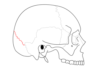
The lambdoid suture is a dense, fibrous connective tissue joint on the posterior aspect of the skull that connects the parietal bones with the occipital bone. It is continuous with the occipitomastoid suture.

<span class="mw-page-title-main">Coronal suture</span> Connective tissue between the parietal bones and the frontal bone of the skull

The coronal suture is a dense, fibrous connective tissue joint that separates the two parietal bones from the frontal bone of the skull.

<span class="mw-page-title-main">Cranial cavity</span> Space inside the skull formed by eight cranial bones known as the neurocranium

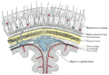
The cranial cavity, also known as intracranial space, is the space within the skull that accommodates the brain. The skull minus the mandible is called the cranium. The cavity is formed by eight cranial bones known as the neurocranium that in humans includes the skull cap and forms the protective case around the brain. The remainder of the skull is called the facial skeleton. Meninges are protective membranes that surround the brain to minimize damage to the brain in the case of head trauma. Meningitis is the inflammation of meninges caused by bacterial or viral infections.

<span class="mw-page-title-main">Superior sagittal sinus</span> Anatomical structure of the brain

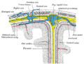
The superior sagittal sinus, within the human head, is an unpaired area along the attached margin of the falx cerebri. It allows blood to drain from the lateral aspects of anterior cerebral hemispheres to the confluence of sinuses. Cerebrospinal fluid drains through arachnoid granulations into the superior sagittal sinus and is returned to venous circulation.

<span class="mw-page-title-main">Bregma</span> Meeting point of the coronal suture and the sagittal suture of the skull

The bregma is the anatomical point on the skull at which the coronal suture is intersected perpendicularly by the sagittal suture.

<span class="mw-page-title-main">Transverse sinuses</span>

The transverse sinuses, within the human head, are two areas beneath the brain which allow blood to drain from the back of the head. They run laterally in a groove along the interior surface of the occipital bone. They drain from the confluence of sinuses to the sigmoid sinuses, which ultimately connect to the internal jugular vein. See diagram : labeled under the brain as "SIN. TRANS.".

<span class="mw-page-title-main">Flat bone</span> Category of bones that function to provide protection or surfaces for muscular attachment

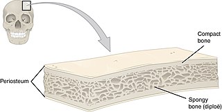
Flat bones are bones whose principal function is either extensive protection or the provision of broad surfaces for muscular attachment. These bones are expanded into broad, flat plates, as in the cranium (skull), the ilium (pelvis), sternum and the rib cage. The flat bones are: the occipital, parietal, frontal, nasal, lacrimal, vomer, hip bone, sternum, ribs, and scapulae.

<span class="mw-page-title-main">Fibrous joint</span> Fixed joints between bones held together by dense, fibrous tissue

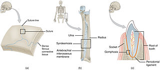
In anatomy, fibrous joints are joints connected by fibrous tissue, consisting mainly of collagen. These are fixed joints where bones are united by a layer of white fibrous tissue of varying thickness. In the skull the joints between the bones are called sutures. Such immovable joints are also referred to as synarthroses.

In arthropod and vertebrate anatomy, the vertex is the highest point of the head.

<span class="mw-page-title-main">Base of skull</span> Inferior area of the skull, composed of the endocranium and lower parts of the skull roof

The base of skull, also known as the cranial base or the cranial floor, is the most inferior area of the skull. It is composed of the endocranium and the lower parts of the calvaria.

<span class="mw-page-title-main">Outline of human anatomy</span> Overview of and topical guide to human anatomy

The following outline is provided as an overview of and topical guide to human anatomy:

The fetal head, from an obstetrical viewpoint, and in particular its size, is important because an essential feature of labor is the adaptation between the fetal head and the maternal bony pelvis. Only a comparatively small part of the head at term is represented by the face. The rest of the head is composed of the firm skull, which is made up of two frontal, two parietal, and two temporal bones, along with the upper portion of the occipital bone and the wings of the sphenoid.

References

  1. Tubbs RS, Bosmia AN, Cohen-Gadol AA (January 2012). "The human calvaria: a review of embryology, anatomy, pathology, and molecular development". Child's Nervous System. 28 (1): 23–31. doi:10.1007/s00381-011-1637-0. PMID   22120469. S2CID   38394369.
  2. Moore KL, Daly AF, Agur AM, eds. (2010). Clinically Oriented Anatomy (6th ed.). Baltimore: Lippincott Williams and Wilkins. p. 1168. ISBN   978-0-7817-7525-0.
  3. Kosaras B, Jakubowski M, Kainz V, Burstein R (July 2009). "Sensory innervation of the calvarial bones of the mouse". The Journal of Comparative Neurology (in French). 515 (3): 331–48. doi:10.1002/cne.22049. PMC   2710390 . PMID   19425099.

Further reading