Lambdoid suture

Last updated
Lambdoid suture
Lambdoid suture.png
Lambdoid suture (shown in red line)
WhiteDesertSkullCropped - Lambdoid suture.png
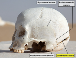
Lambdoid suture (labeled at bottom right)
Details
Part of skull
Nerve supraorbital nerve
Identifiers
Latin sutura lambdoidea
TA98 A03.1.02.004
TA2 1577
FMA 52933
Anatomical terms of bone

The lambdoid suture (or lambdoidal suture) is a dense, fibrous connective tissue joint on the posterior aspect of the skull that connects the parietal bones with the occipital bone. It is continuous with the occipitomastoid suture.

Contents

Structure

The lambdoid suture is between the paired parietal bones and the occipital bone of the skull. It runs from the asterion on each side.

Nerve supply

The lambdoid suture may be supplied by a branch of the supraorbital nerve, a branch of the frontal branch of the trigeminal nerve. [1] [2]

Clinical significance

At birth, the bones of the skull do not meet. If certain bones of the skull grow too fast, then craniosynostosis (premature closure of the sutures) may occur. This can result in skull deformities. If the lambdoid suture closes too soon on one side, the skull will appear twisted and asymmetrical, a condition called "plagiocephaly". Plagiocephaly refers to the shape and not the condition. The condition is craniosynostosis.[ citation needed ]

The lambdoid suture can be damaged by a fall backward. [2]

Etymology

The lambdoid suture is named due to its uppercase lambda-like shape.

Additional images

Related Research Articles

<span class="mw-page-title-main">Skull</span> Bony structure that forms the head in vertebrates

The skull is a bone protective cavity for the brain. The skull is composed of four types of bone i.e., cranial bones, facial bones, ear ossicles and hyoid bone, however two parts are more prominent: the cranium and the mandible. In humans, these two parts are the neurocranium (braincase) and the viscerocranium that includes the mandible as its largest bone. The skull forms the anterior-most portion of the skeleton and is a product of cephalisation—housing the brain, and several sensory structures such as the eyes, ears, nose, and mouth. In humans, these sensory structures are part of the facial skeleton.

<span class="mw-page-title-main">Occipital bone</span> Skull bone, lower back of cranium

The occipital bone is a cranial dermal bone and the main bone of the occiput. It is trapezoidal in shape and curved on itself like a shallow dish. The occipital bone overlies the occipital lobes of the cerebrum. At the base of the skull in the occipital bone, there is a large oval opening called the foramen magnum, which allows the passage of the spinal cord.

<span class="mw-page-title-main">Craniosynostosis</span> Premature fusion of bones in the skull

Craniosynostosis is a condition in which one or more of the fibrous sutures in a young infant's skull prematurely fuses by turning into bone (ossification), thereby changing the growth pattern of the skull. Because the skull cannot expand perpendicular to the fused suture, it compensates by growing more in the direction parallel to the closed sutures. Sometimes the resulting growth pattern provides the necessary space for the growing brain, but results in an abnormal head shape and abnormal facial features. In cases in which the compensation does not effectively provide enough space for the growing brain, craniosynostosis results in increased intracranial pressure leading possibly to visual impairment, sleeping impairment, eating difficulties, or an impairment of mental development combined with a significant reduction in IQ.

<span class="mw-page-title-main">Posterior cranial fossa</span> Area of the cranium containing the brainstem and cerebellum

The posterior cranial fossa is the part of the cranial cavity located between the foramen magnum, and tentorium cerebelli. It is formed by the sphenoid bones, temporal bones, and occipital bone. It lodges the cerebellum, and parts of the brainstem.

<span class="mw-page-title-main">Superior orbital fissure</span> Foramen in the skull allowing for passage of cranial nerves

The superior orbital fissure is a foramen or cleft of the skull between the lesser and greater wings of the sphenoid bone. It gives passage to multiple structures, including the oculomotor nerve, trochlear nerve, ophthalmic nerve, abducens nerve, ophthalmic veins, and sympathetic fibres from the cavernous plexus.

<span class="mw-page-title-main">Sagittal suture</span> Midline joint between the parietal bones of the skull

The sagittal suture, also known as the interparietal suture and the sutura interparietalis, is a dense, fibrous connective tissue joint between the two parietal bones of the skull. The term is derived from the Latin word sagitta, meaning arrow.

<span class="mw-page-title-main">Coronal suture</span> Connective tissue between the parietal bones and the frontal bone of the skull

The coronal suture is a dense, fibrous connective tissue joint that separates the two parietal bones from the frontal bone of the skull.

<span class="mw-page-title-main">Foramen spinosum</span> Hole in the sphenoid bone of the skull

The foramen spinosum is a small open hole in the greater wing of the sphenoid bone that gives passage to the middle meningeal artery and vein, and the meningeal branch of the mandibular nerve.

<span class="mw-page-title-main">Occipitofrontalis muscle</span> Facial muscle helping to create facial expressions

The occipitofrontalis muscle is a muscle which covers parts of the skull. It consists of two parts or bellies: the occipital belly, near the occipital bone, and the frontal belly, near the frontal bone. It is supplied by the supraorbital artery, the supratrochlear artery, and the occipital artery. It is innervated by the facial nerve. In humans, the occipitofrontalis helps to create facial expressions.

<span class="mw-page-title-main">Foramen rotundum</span> Hole in the sphenoid bone of the skull

The foramen rotundum is a circular hole in the sphenoid bone of the skull. It connects the middle cranial fossa and the pterygopalatine fossa. It allows for the passage of the maxillary nerve (V2), a branch of the trigeminal nerve.

<span class="mw-page-title-main">Jugular foramen</span> Opening in the base of the skull allowing many structures to pass

A jugular foramen is one of the two large foramina (openings) in the base of the skull, located behind the carotid canal. It is formed by the temporal bone and the occipital bone. It allows many structures to pass, including the inferior petrosal sinus, three cranial nerves, the sigmoid sinus, and meningeal arteries.

<span class="mw-page-title-main">Supratrochlear nerve</span> Nerve of the forehead

The supratrochlear nerve is a branch of the frontal nerve, itself a branch of the ophthalmic nerve (CN V1) from the trigeminal nerve (CN V). It provides sensory innervation to the skin of the forehead and the upper eyelid.

<span class="mw-page-title-main">Lateral sacral artery</span> Artery in the pelvis

The lateral sacral arteries is an artery in the pelvis that arises from the posterior division of the internal iliac artery. It later splits into two smaller branches, a superior and an inferior.

<span class="mw-page-title-main">Mastoid part of the temporal bone</span> Back part of the sides of the skull base

The mastoid part of the temporal bone is the posterior (back) part of the temporal bone, one of the bones of the skull. Its rough surface gives attachment to various muscles and it has openings for blood vessels. From its borders, the mastoid part articulates with two other bones.

<span class="mw-page-title-main">Middle cranial fossa</span>

The middle cranial fossa is formed by the sphenoid bones, and the temporal bones. It lodges the temporal lobes, and the pituitary gland. It is deeper than the anterior cranial fossa, is narrow medially and widens laterally to the sides of the skull. It is separated from the posterior cranial fossa by the clivus and the petrous crest.

<span class="mw-page-title-main">Posterior ethmoidal nerve</span> Nerve of the orbit around the eye

The posterior ethmoidal nerve is a nerve of the head. It is a branch of the nasociliary nerve (itself a branch of the ophthalmic nerve (CN V1)). It provides sensory innervation to the sphenoid sinus and ethmoid sinus, and part of the dura mater in the anterior cranial fossa.

<span class="mw-page-title-main">Medial pterygoid nerve</span> Nerve of the head

The medial pterygoid nerve (nerve to medial pterygoid, or internal pterygoid nerve) is a nerve of the head. It is a branch of the mandibular nerve (CN V3). It supplies the medial pterygoid muscle, the tensor veli palatini muscle, and the tensor tympani muscle.

<span class="mw-page-title-main">Calvaria (skull)</span> Top part of the skull

The calvaria is the top part of the skull. It is the superior part of the neurocranium and covers the cranial cavity containing the brain. It forms the main component of the skull roof.

<span class="mw-page-title-main">Superior auricular muscle</span> Muscle that draws the auricle of the outer ear upwards

The superior auricular muscle is a muscle above the auricle of the outer ear. It originates from the epicranial aponeurosis, and inserts into the upper part of the medial surface of the auricle. It draws the auricle upwards.

<span class="mw-page-title-main">Anterior auricular muscle</span> Muscle connectingthe epicranial aponeurosis to the helix of the ear

The anterior auricular muscle, the smallest of the three auricular muscles, is thin and fan-shaped, and its fibers are pale and indistinct. It arises from the lateral edge of the epicranial aponeurosis, and its fibers converge to be inserted into a projection on the front of the helix.

References

  1. Barral, Jean-Pierre; Croibier, Alain (2009-01-01). "2 - Characteristics of cranial nerves". Manual Therapy for the Cranial Nerves. Churchill Livingstone. pp. 7–14. doi:10.1016/b978-0-7020-3100-7.50005-7. ISBN   978-0-7020-3100-7.{{cite book}}: CS1 maint: date and year (link)
  2. 1 2 Barral, Jean-Pierre; Croibier, Alain (2009-01-01). "15 - Ophthalmic nerve". Manual Therapy for the Cranial Nerves. Churchill Livingstone. pp. 115–128. doi:10.1016/b978-0-7020-3100-7.50018-5. ISBN   978-0-7020-3100-7.{{cite book}}: CS1 maint: date and year (link)