The maxilla in vertebrates is the upper fixed bone of the jaw formed from the fusion of two maxillary bones. In humans, the upper jaw includes the hard palate in the front of the mouth. The two maxillary bones are fused at the intermaxillary suture, forming the anterior nasal spine. This is similar to the mandible, which is also a fusion of two mandibular bones at the mandibular symphysis. The mandible is the movable part of the jaw.

In the human skull, the zygomatic bone is a paired irregular bone which articulates with the maxilla, the temporal bone, the sphenoid bone and the frontal bone. It is situated at the upper and lateral part of the face and forms the prominence of the cheek, part of the lateral wall and floor of the orbit, and parts of the temporal fossa and the infratemporal fossa. It presents a malar and a temporal surface; four processes, and four borders.
The occipital bone is a cranial dermal bone and the main bone of the occiput. It is trapezoidal in shape and curved on itself like a shallow dish. The occipital bone overlies the occipital lobes of the cerebrum. At the base of skull in the occipital bone, there is a large oval opening called the foramen magnum, which allows the passage of the spinal cord.

The parietal bones are two bones in the skull which, when joined together at a fibrous joint, form the sides and roof of the cranium. In humans, each bone is roughly quadrilateral in form, and has two surfaces, four borders, and four angles. It is named from the Latin paries (-ietis), wall.

The temporal bones are situated at the sides and base of the skull, and lateral to the temporal lobes of the cerebral cortex.

In anatomy, the zygomatic arch, or cheek bone, is a part of the skull formed by the zygomatic process of the temporal bone and the temporal process of the zygomatic bone, the two being united by an oblique suture ; the tendon of the temporal muscle passes medial to the arch, to gain insertion into the coronoid process of the mandible (jawbone).

The temporal fossa is a fossa on the side of the skull bounded by the temporal lines and terminating below the level of the zygomatic arch.

The condyloid process or condylar process is the process on the human mandible and some other species' mandibles that ends in a condyle, the mandibular condyle. It is thicker than the coronoid process of the mandible and consists of two portions: the condyle and the constricted portion which supports it, the neck.

The zygomaticotemporal nerve (zygomaticotemporal branch, temporal branch) is a small nerve of the face. It is derived from the zygomatic nerve, a branch of the maxillary nerve (CN V2). It is distributed to the skin of the side of the forehead. It communicates with the facial nerve and with the auriculotemporal branch of the mandibular nerve.

The squamous part of temporal bone, or temporal squama, forms the front and upper part of the temporal bone, and is scale-like, thin, and translucent.

There are two surfaces of the squamous part of the frontal bone: the external surface, and the internal surface.

The squamosal suture, or squamous suture, arches backward from the pterion and connects the temporal squama with the lower border of the parietal bone: this suture is continuous behind with the short, nearly horizontal parietomastoid suture, which unites the mastoid process of the temporal with the region of the mastoid angle of the parietal bone. The term parietotemporal suture may refer to both of these sutures or exclusively to the parietomastoid suture and its use is, therefore, best avoided.

The sphenozygomatic suture is the cranial suture between the sphenoid bone and the zygomatic bone.

The Sphenoparietal suture is the cranial suture between the sphenoid bone and the parietal bone. It is one of the sutures that comprises the pterion.

The occipitomastoid suture or occipitotemporal suture is the cranial suture between the occipital bone and the mastoid portion of the temporal bone.
The Sphenosquamosal suture is a cranial suture between the sphenoid bone and the squama of the temporal bone.

The zygomatico-orbital foramina are two canals in the skull, that allow nerves to pass through. The orifices are seen on the orbital process of the zygomatic bone.
The zygomatic processes are three processes (protrusions) from other bones of the skull which each articulate with the zygomatic bone. The three processes are:
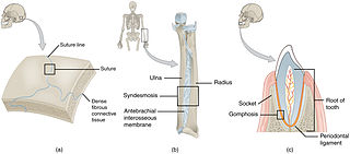
In anatomy, fibrous joints are joints connected by fibrous tissue, consisting mainly of collagen. These are fixed joints where bones are united by a layer of white fibrous tissue of varying thickness. In the skull the joints between the bones are called sutures. Such immovable joints are also referred to as synarthroses.
In human anatomy, the mandible's coronoid process is a thin, triangular eminence, which is flattened from side to side and varies in shape and size. Its anterior border is convex and is continuous below with the anterior border of the ramus. Its posterior border is concave and forms the anterior boundary of the mandibular notch. The lateral surface is smooth, and affords insertion to the temporalis and masseter muscles. Its medial surface gives insertion to the temporalis, and presents a ridge which begins near the apex of the process and runs downward and forward to the inner side of the last molar tooth.
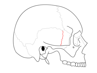
 Zygomaticotemporal suture (blue circle) and position of two bones.
 Position of zygomaticotemporal suture (red). Animation.
 Cross section (temporal bones removed). Animation.
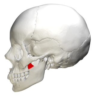
 Still image (red)
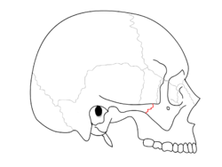
 Side view of the skull. (Zygomaticotemporal suture is suture between zygomatic bone, at left in white, and temporal bone, at center in pink.)