The skull is a bone protective cavity for the brain. The skull is composed of four types of bone i.e., cranial bones, facial bones, ear ossicles and hyoid bone, however two parts are more prominent: the cranium and the mandible. In humans, these two parts are the neurocranium (braincase) and the viscerocranium that includes the mandible as its largest bone. The skull forms the anterior-most portion of the skeleton and is a product of cephalisation—housing the brain, and several sensory structures such as the eyes, ears, nose, and mouth. In humans, these sensory structures are part of the facial skeleton.

The maxilla in vertebrates is the upper fixed bone of the jaw formed from the fusion of two maxillary bones. In humans, the upper jaw includes the hard palate in the front of the mouth. The two maxillary bones are fused at the intermaxillary suture, forming the anterior nasal spine. This is similar to the mandible, which is also a fusion of two mandibular bones at the mandibular symphysis. The mandible is the movable part of the jaw.

The occipital bone is a cranial dermal bone and the main bone of the occiput. It is trapezoidal in shape and curved on itself like a shallow dish. The occipital bone overlies the occipital lobes of the cerebrum. At the base of the skull in the occipital bone, there is a large oval opening called the foramen magnum, which allows the passage of the spinal cord.

The frontal bone or sincipital bone is a bone in the human skull. The bone consists of two portions. These are the vertically oriented squamous part, and the horizontally oriented orbital part, making up the bony part of the forehead, part of the bony orbital cavity holding the eye, and part of the bony part of the nose respectively. The name comes from the Latin word frons.

The parietal bones are two bones in the skull which, when joined at a fibrous joint, form the sides and roof of the cranium. In humans, each bone is roughly quadrilateral in form, and has two surfaces, four borders, and four angles. It is named from the Latin paries (-ietis), wall.

In anatomy, the zygomatic arch, or cheek bone, is a part of the skull formed by the zygomatic process of the temporal bone and the temporal process of the zygomatic bone, the two being united by an oblique suture ; the tendon of the temporal muscle passes medial to the arch, to gain insertion into the coronoid process of the mandible (jawbone).

The sagittal suture, also known as the interparietal suture and the sutura interparietalis, is a dense, fibrous connective tissue joint between the two parietal bones of the skull. The term is derived from the Latin word sagitta, meaning arrow.

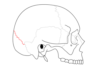
The lambdoid suture is a dense, fibrous connective tissue joint on the posterior aspect of the skull that connects the parietal bones with the occipital bone. It is continuous with the occipitomastoid suture.

The coronal suture is a dense, fibrous connective tissue joint that separates the two parietal bones from the frontal bone of the skull.

Wormian bones, also known as intrasutural bones or sutural bones, are extra bone pieces that can occur within a suture (joint) in the skull. These are irregular isolated bones that can appear in addition to the usual centres of ossification of the skull and, although unusual, are not rare. They occur most frequently in the course of the lambdoid suture, which is more tortuous than other sutures. They are also occasionally seen within the sagittal and coronal sutures. A large wormian bone at lambda is often called an Inca bone , due to the relatively high frequency of occurrence in Peruvian mummies. Another specific Wormian bone, the pterion ossicle, sometimes exists between the sphenoidal angle of the parietal bone and the great wing of the sphenoid bone. They tend to vary in size and can be found on either side of the skull. Usually, not more than several are found in a single individual, but more than one hundred have been once found in the skull of a hydrocephalic adult.

The sphenozygomatic suture is the cranial suture between the sphenoid bone and the zygomatic bone.

The sphenoparietal suture is the cranial suture between the sphenoid bone and the parietal bone. It is one of the sutures that comprises the pterion.

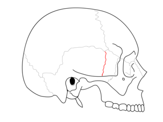
The zygomaticofrontal suture is the cranial suture between the zygomatic bone and the frontal bone. The suture can be palpated just lateral to the eye.

The zygomaticotemporal suture is the cranial suture between the zygomatic bone and the temporal bone. This is part of the zygomatic arch. Movement at the suture decreases with development during aging. It has a complex internal structure.

The occipitomastoid suture or occipitotemporal suture is the cranial suture between the occipital bone and the mastoid portion of the temporal bone.

The sphenosquamosal suture is a cranial suture between the sphenoid bone and the squama of the temporal bone.

The pterion is the region where the frontal, parietal, temporal, and sphenoid bones join. It is located on the side of the skull, just behind the temple. It is also considered to be the weakest part of the skull, which makes it clinically significant, as if there is a fracture around the pterion it could be accompanied by an epidural hematoma.

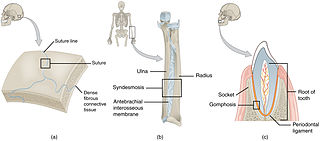
In anatomy, fibrous joints are joints connected by fibrous tissue, consisting mainly of collagen. These are fixed joints where bones are united by a layer of white fibrous tissue of varying thickness. In the skull, the joints between the bones are called sutures. Such immovable joints are also referred to as synarthroses.
In arthropod and vertebrate anatomy, the vertex is the highest point of the head.

In anatomy, a suture is a fairly rigid joint between two or more hard elements of an organism, with or without significant overlap of the elements.


