The skull is a bone protective cavity for the brain. The skull is composed of four types of bone i.e., cranial bones, facial bones, ear ossicles and hyoid bone, however two parts are more prominent: the cranium and the mandible. In humans, these two parts are the neurocranium (braincase) and the viscerocranium that includes the mandible as its largest bone. The skull forms the anterior-most portion of the skeleton and is a product of cephalisation—housing the brain, and several sensory structures such as the eyes, ears, nose, and mouth. In humans, these sensory structures are part of the facial skeleton.

A fontanelle is an anatomical feature of the infant human skull comprising soft membranous gaps (sutures) between the cranial bones that make up the calvaria of a fetus or an infant. Fontanelles allow for stretching and deformation of the neurocranium both during birth and later as the brain expands faster than the surrounding bone can grow. Premature complete ossification of the sutures is called craniosynostosis.

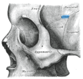
The sphenoid bone is an unpaired bone of the neurocranium. It is situated in the middle of the skull towards the front, in front of the basilar part of the occipital bone. The sphenoid bone is one of the seven bones that articulate to form the orbit. Its shape somewhat resembles that of a butterfly or bat with its wings extended.

The occipital bone is a cranial dermal bone and the main bone of the occiput. It is trapezoidal in shape and curved on itself like a shallow dish. The occipital bone overlies the occipital lobes of the cerebrum. At the base of the skull in the occipital bone, there is a large oval opening called the foramen magnum, which allows the passage of the spinal cord.

The frontal bone or sincipital bone is a bone in the human skull. The bone consists of two portions. These are the vertically oriented squamous part, and the horizontally oriented orbital part, making up the bony part of the forehead, part of the bony orbital cavity holding the eye, and part of the bony part of the nose respectively. The name comes from the Latin word frons.
The parietal bones are two bones in the skull which, when joined at a fibrous joint, form the sides and roof of the cranium. In humans, each bone is roughly quadrilateral in form, and has two surfaces, four borders, and four angles. It is named from the Latin paries (-ietis), wall.

The temporal bones are situated at the sides and base of the skull, and lateral to the temporal lobes of the cerebral cortex.

The middle meningeal artery is typically the third branch of the first portion of the maxillary artery. After branching off the maxillary artery in the infratemporal fossa, it runs through the foramen spinosum to supply the dura mater and the calvaria. The middle meningeal artery is the largest of the three (paired) arteries that supply the meninges, the others being the anterior meningeal artery and the posterior meningeal artery.
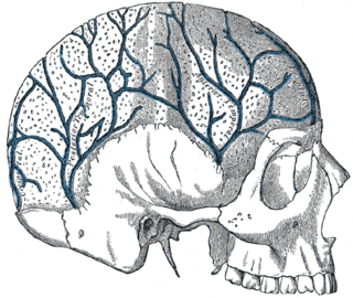
The diploic veins are large, thin-walled valveless veins that channel in the diploë between the inner and outer layers of the cortical bone in the skull, first identified in dogs by the anatomist Guillaume Dupuytren. A single layer of endothelium lines these veins supported by elastic tissue. They develop fully by the age of two years. The diploic veins drain this area into the dural venous sinuses. The four major trunks of the diploic veins found on each side of the head are frontal, anterior temporal, posterior temporal, and occipital diploic veins. They tend to be symmetrical, with the same pattern of large veins on each side of the skull. It has been suggested that the venous patterns they form resemble fingerprints in their individuality.

The foramen spinosum is a small open hole in the greater wing of the sphenoid bone that gives passage to the middle meningeal artery and vein, and the meningeal branch of the mandibular nerve.

The cranial cavity, also known as intracranial space, is the space within the skull that accommodates the brain. The skull minus the mandible is called the cranium. The cavity is formed by eight cranial bones known as the neurocranium that in humans includes the skull cap and forms the protective case around the brain. The remainder of the skull is called the facial skeleton. Meninges are protective membranes that surround the brain to minimize damage to the brain in the case of head trauma. Meningitis is the inflammation of meninges caused by bacterial or viral infections.

Wormian bones, also known as intrasutural bones or sutural bones, are extra bone pieces that can occur within a suture (joint) in the skull. These are irregular isolated bones that can appear in addition to the usual centres of ossification of the skull and, although unusual, are not rare. They occur most frequently in the course of the lambdoid suture, which is more tortuous than other sutures. They are also occasionally seen within the sagittal and coronal sutures. A large wormian bone at lambda is often called an Inca bone , due to the relatively high frequency of occurrence in Peruvian mummies. Another specific Wormian bone, the pterion ossicle, sometimes exists between the sphenoidal angle of the parietal bone and the great wing of the sphenoid bone. They tend to vary in size and can be found on either side of the skull. Usually, not more than several are found in a single individual, but more than one hundred have been once found in the skull of a hydrocephalic adult.

The squamous part of temporal bone, or temporal squama, forms the front and upper part of the temporal bone, and is scale-like, thin, and translucent.

The basilar part of the occipital bone extends forward and upward from the foramen magnum, and presents in front an area more or less quadrilateral in outline.

The middle cranial fossa is formed by the sphenoid bones, and the temporal bones. It lodges the temporal lobes, and the pituitary gland. It is deeper than the anterior cranial fossa, is narrow medially and widens laterally to the sides of the skull. It is separated from the posterior cranial fossa by the clivus and the petrous crest.

The sphenozygomatic suture is the cranial suture between the sphenoid bone and the zygomatic bone.

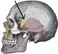
The pterion is the region where the frontal, parietal, temporal, and sphenoid bones join. It is located on the side of the skull, just behind the temple. It is also considered to be the weakest part of the skull, which makes it clinically significant, as if there is a fracture around the pterion it could be accompanied by an epidural hematoma.
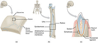
In anatomy, fibrous joints are joints connected by fibrous tissue, consisting mainly of collagen. These are fixed joints where bones are united by a layer of white fibrous tissue of varying thickness. In the skull, the joints between the bones are called sutures. Such immovable joints are also referred to as synarthroses.

The sphenoparietal sinus is a paired dural venous sinus situated along the posterior edge of the lesser wing of either sphenoid bone. It drains into the cavernous sinus.

In human anatomy, the neurocranium, also known as the braincase, brainpan, or brain-pan, is the upper and back part of the skull, which forms a protective case around the brain. In the human skull, the neurocranium includes the calvaria or skullcap. The remainder of the skull is the facial skeleton.