Anterior pituitary

Last updated
Anterior pituitary gland
Gray1181.png
Median sagittal through the hypophysis of an adult monkey. Semidiagrammatic.
Details
Precursor Oral mucosa (Rathke's pouch)
Artery superior hypophyseal
Vein hypophyseal
Identifiers
Latin lobus anterior hypophysis
MeSH D010903
NeuroNames 407
NeuroLex ID birnlex_1581
TA98 A11.1.00.002
TA2 3855
FMA 74627
Anatomical terminology

A major organ of the endocrine system, the anterior pituitary (also called the adenohypophysis or pars anterior) is the glandular, anterior lobe that together with the posterior lobe (posterior pituitary, or the neurohypophysis) makes up the pituitary gland (hypophysis) which, in humans, is located at the base of the brain, protruding off the bottom of the hypothalamus.

Contents

The anterior pituitary regulates several physiological processes, including stress, growth, reproduction, and lactation. Proper functioning of the anterior pituitary and of the organs it regulates can often be ascertained via blood tests that measure hormone levels.

Structure

The anterior pituitary complex 1808 The Anterior Pituitary Complex.jpg
The anterior pituitary complex

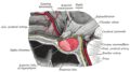
The pituitary gland sits in a protective bony enclosure called the sella turcica (Turkish chair/saddle). It is composed of three lobes: the anterior, intermediate, and posterior lobes. In many animals, these lobes are distinct. However, in humans, the intermediate lobe is but a few cell layers thick and indistinct; as a result, it is often considered part of the anterior pituitary. In all animals, the fleshy, glandular anterior pituitary is distinct from the neural composition of the posterior pituitary.[ citation needed ]

The anterior pituitary is composed of three regions:

Pars distalis

The pars distalis (distal part) comprises the majority of the anterior pituitary and is where the bulk of pituitary hormone production occurs. The pars distalis contains two types of cells, including chromophobe cells and chromophil cells. [1] The chromophils can be further divided into acidophils (alpha cells) and basophils (beta cells). [1] These cells all together produce hormones of the anterior pituitary and release them into the blood stream.[ citation needed ]

Nota bene: The terms "basophil" and "acidophil" are used by some books, whereas others prefer not to use these terms. This is due to the possible confusion with white blood cells, where one may also find basophils and acidophils.

Pars tuberalis
The pars tuberalis (tubular part) forms a part of the sheath extending up from the pars distalis, which joins with the pituitary stalk (also known as the infundibular stalk or infundibulum), arising from the posterior lobe. (The pituitary stalk connects the hypothalamus to the posterior pituitary.) The function of the pars tuberalis is poorly understood. However, it has been seen to be important in receiving the endocrine signal in the form of TSHB (a β subunit of TSH), informing the pars tuberalis of the photoperiod (length of day). The expression of this subunit is regulated by the secretion of melatonin in response to light information transmitted to the pineal gland. [2] [3] Earlier studies have shown localization of melatonin receptors in this region. [4]
Pars intermedia
The pars intermedia (intermediate part) sits between the pars distalis and the posterior pituitary, forming the boundary between the anterior and posterior pituitaries. It is very small and indistinct in humans.

Development

The anterior pituitary is derived from the ectoderm, more specifically from that of Rathke's pouch, part of the developing hard palate in the embryo. Rathke's pouch is also ectodermal in origin.

The pouch eventually loses its connection with the pharynx, giving rise to the anterior pituitary. The anterior wall of Rathke's pouch proliferates, filling most of the pouch to form the pars distalis and the pars tuberalis. The posterior wall of the anterior pituitary forms the pars intermedia. Its formation from the soft tissues of the upper palate contrasts with the posterior pituitary, which originates from neuroectoderm. [5]

Function

The anterior pituitary contains five types of endocrine cell, and they are defined by the hormones they secrete: somatotropes (GH); lactotropes (PRL); gonadotropes (LH and FSH); corticotropes (ACTH) and thyrotropes (TSH). [6] It also contains non-endocrine folliculostellate cells which are thought to stimulate and support the endocrine cell populations.

Hormones secreted by the anterior pituitary are trophic hormones (Greek: trophe, "nourishment"). Trophic hormones directly affect growth either as hyperplasia or hypertrophy on the tissue it is stimulating. Tropic hormones are named for their ability to act directly on target tissues or other endocrine glands to release hormones, causing numerous cascading physiological responses. [5]

HormoneOther namesSymbol(s)StructureSecretory cellsStainingTargetEffect
Adrenocorticotropic hormone CorticotropinACTH Polypeptide Corticotrophs Basophil Adrenal gland Secretion of glucocorticoid, mineralocorticoid and androgens
Thyroid-stimulating hormone ThyrotropinTSH Glycoprotein Thyrotrophs Basophil Thyroid gland Secretion of thyroid hormones
Follicle-stimulating hormone -FSHGlycoprotein Gonadotrophs Basophil Gonads Growth of reproductive system
Luteinizing hormone LutropinLH, ICSHGlycoprotein Gonadotrophs Basophil Gonads Sex hormone production
Growth hormone SomatotropinGH, STHPolypeptide Somatotrophs Acidophil Liver, adipose tissue Promotes growth; lipid and carbohydrate metabolism
Prolactin LactotropinPRLPolypeptide Lactotrophs Acidophil Ovaries, mammary glands, testes, prostate Secretion of estrogens/progesterone; lactation; spermatogenesis; prostatic hyperplasia TSH and ACTH secretion

[7] [8]

Role in the endocrine system

Hypothalamic control

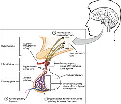
Hormone secretion from the anterior pituitary gland is regulated by hormones secreted by the hypothalamus. Neuroendocrine cells in the hypothalamus project axons to the median eminence, at the base of the brain. At this site, these cells can release substances into small blood vessels that travel directly to the anterior pituitary gland (the hypothalamo-hypophyseal portal vessels).

Other mechanisms

Aside from hypothalamic control of the anterior pituitary, other systems in the body have been shown to regulate the anterior pituitary's function. GABA can either stimulate or inhibit the secretion of luteinizing hormone (LH) and growth hormone (GH) and can stimulate the secretion of thyroid-stimulating hormone (TSH). Prostaglandins are now known to inhibit adrenocorticotropic hormone (ACTH) and also to stimulate TSH, GH and LH release. [9] Clinical evidence supports the experimental findings of the excitatory and inhibitory effects GABA has on GH secretion, dependent on GABA's site of action within the hypothalamic-pituitary axis. [10]

Effects of the anterior pituitary

Thermal homeostasis

The homeostatic maintenance of the anterior pituitary is crucial to our physiological well being. Increased plasma levels of TSH induce hyperthermia through a mechanism involving increased metabolism and cutaneous vasodilation. Increased levels of LH also result in hypothermia but through a decreased metabolism action. ACTH increase metabolism and induce cutaneous vasoconstriction, increased plasma levels also result in hyperthermia and prolactin decreases with decreasing temperature values. follicle-stimulating hormone (FSH) also may cause hypothermia if increased beyond homeostatic levels through an increased metabolic mechanism only. [11]

Gonadal function

Gonadotropes, primarily luteinising hormone (LH) secreted from the anterior pituitary stimulates the ovulation cycle in female mammals, whilst in the males, LH stimulates the synthesis of androgen which drives the ongoing will to mate together with a constant production of sperm. [5]

HPA axis

Main article Hypothalamic-pituitary-adrenal axis

The anterior pituitary plays a role in stress response. Corticotropin releasing hormone (CRH) from the hypothalamus stimulates ACTH release in a cascading effect that ends with the production of glucocorticoids from the adrenal cortex. [5]

Behavioral effects

Development
The release of GH, LH, and FSH are required for correct human development, including gonadal development. [12]
Breast-feeding
Release of the hormone prolactin is essential for lactation. [12]
Stress
Operating through the hypothalamic-pituitary-adrenal axis (HPA), the anterior pituitary gland has a large role in the neuroendocrine system's stress response. Stress induces a release of corticotropin-releasing hormone (CRH) and vasopressin from the hypothalamus, which activates the release of adrenocorticotropic hormone (ACTH) from the anterior pituitary gland. Then, this acts on the adrenal cortex to produce glucocorticoids such as cortisol. These glucocorticoids act back on the anterior pituitary gland and the hypothalamus with negative feedback to slow the production of CRH and ACTH. [13] [14] Increased cortisol under stress conditions can cause the following: metabolic effects (mobilization of glucose, fatty acids, and amino acids), bone re-absorption (calcium mobilization), activation of the sympathetic nervous system response (fight or flight), anti-inflammatory effects, and inhibition of reproduction/growth. [12] When the anterior pituitary gland is removed (hypophysectomy) in rats, their avoidance learning mechanisms were slowed, but injections of ACTH restored their performance. [12] In addition, stress may delay the release of reproductive hormones such as luteinizing hormone (LH) and follicle-stimulating hormone (FSH). [15] This shows that the anterior pituitary gland is involved in behavioral functions as well as being part of a larger pathway for stress responses. It is also known that (HPA) hormones are related to certain skin diseases and skin homeostasis. There is evidence linking hyperactivity of HPA hormones to stress-related skin diseases and skin tumors. [16]
Aging
Operating through the hypothalamic-pituitary-gonadal axis, the anterior pituitary gland also affects the reproductive system. The hypothalamus releases gonadotropin-releasing hormone (GnRH), which stimulates the release of luteinizing hormone (LH) and follicle-stimulating hormone. Then the gonads produce estrogen and testosterone. The decrease in release of gonadotropins (LH and FSH) caused by normal aging may be responsible for impotence [12] [15] and frailty [17] in elderly men because of the eventual decrease in production of testosterone. This lower level of testosterone can have other effects, such as reduced libido, well-being and mood, muscle and bone strength, and metabolism. [15]
Tactile responding
It has been shown that infant mice who were stroked with a paintbrush (simulating motherly care) had more release and binding of growth hormone (GH) from the anterior pituitary gland. [12]
Circadian rhythms
Light information received by the eyes is transmitted to the pineal gland via the circadian pacemaker (the suprachiasmatic nucleus). Diminishing light stimulates the release of melatonin from the pineal gland which can also affect the secretion levels in the hypothalamic-pituitary-gonadal axis. [12] Melatonin can lower levels of LH and FSH, which will decrease levels of estrogen and testosterone. In addition, melatonin may affect production of prolactin. [18]

Clinical significance

Increased activity

Hyperpituitarism is the condition where the pituitary secretes excessive amounts of hormones. This hypersecretion often results in the formation of a pituitary adenoma (tumour), which are benign apart from a tiny fraction. There are mainly three types of anterior pituitary tumors and their associated disorders. For example, acromegaly results from excessive secretion of growth hormone (GH) often being released by a pituitary adenoma. This disorder can cause disfigurement and possibly death [19] and can lead to gigantism, a hormone disorder shown in "giants" such as André the Giant, where it occurs before the epiphyseal plates in bones close in puberty. [12] The most common type of pituitary tumour is a prolactinoma which hypersecretes prolactin. [20] A third type of pituitary adenoma secretes excess ACTH, which in turn, causes an excess of cortisol to be secreted and is the cause of Cushing's disease. [12]

Decreased activity

Hypopituitarism is characterized by a decreased secretion of hormones released by the anterior pituitary. For example, hypo-secretion of GH prior to puberty can be a cause of dwarfism. In addition, secondary adrenal insufficiency can be caused by hypo-secretion of ACTH which, in turn, does not signal the adrenal cortex to produce a sufficient amount of cortisol. This is a life-threatening condition. Hypopituitarism could be caused by the destruction or removal of the anterior pituitary tissue through traumatic brain injury, tumor, tuberculosis, or syphilis, among other causes. This disorder used to be referred to as Simmonds' disease but now according to the Diseases Database it is called Sheehan syndrome. [21] If the hypopituitarism is caused by the blood loss associated with childbirth, the disorder is referred to as Sheehan syndrome.

History

Etymology

The anterior pituitary is also known as the adenohypophysis, meaning "glandular undergrowth", from the Greek adeno- ("gland"), hypo ("under"), and physis ("growth").

Additional images

See also

Related Research Articles

<span class="mw-page-title-main">Endocrine system</span> Hormone-producing glands of a body

The endocrine system is a messenger system in an organism comprising feedback loops of hormones that are released by internal glands directly into the circulatory system and that target and regulate distant organs. In vertebrates, the hypothalamus is the neural control center for all endocrine systems.

<span class="mw-page-title-main">Pituitary gland</span> Endocrine gland at the base of the brain

The pituitary gland is an endocrine gland in vertebrates. In humans, the pituitary gland is located at the base of the brain, protruding off the bottom of the hypothalamus. The human pituitary gland is oval shaped, about the size of a chickpea, and weighs 0.5 grams (0.018 oz) on average.

<span class="mw-page-title-main">Adrenocorticotropic hormone</span> Pituitary hormone

Adrenocorticotropic hormone is a polypeptide tropic hormone produced by and secreted by the anterior pituitary gland. It is also used as a medication and diagnostic agent. ACTH is an important component of the hypothalamic-pituitary-adrenal axis and is often produced in response to biological stress. Its principal effects are increased production and release of cortisol and androgens by the cortex and medulla of the adrenal gland, respectively. ACTH is also related to the circadian rhythm in many organisms.

<span class="mw-page-title-main">Hypothalamus</span> Area of the brain below the thalamus

The hypothalamus is a small part of the brain that contains a number of nuclei with a variety of functions. One of the most important functions is to link the nervous system to the endocrine system via the pituitary gland. The hypothalamus is located below the thalamus and is part of the limbic system. It forms the ventral part of the diencephalon. All vertebrate brains contain a hypothalamus. In humans, it is the size of an almond.

<span class="mw-page-title-main">Hypothalamic–pituitary–adrenal axis</span> Set of physiological feedback interactions

The hypothalamic–pituitary–adrenal axis is a complex set of direct influences and feedback interactions among three components: the hypothalamus, the pituitary gland, and the adrenal glands. These organs and their interactions constitute the HPA axis.

Tropic hormones are hormones that have other endocrine glands as their target. Most tropic hormones are produced and secreted by the anterior pituitary. The hypothalamus secretes tropic hormones that target the anterior pituitary, and the thyroid gland secretes thyroxine, which targets the hypothalamus and therefore can be considered a tropic hormone.

<span class="mw-page-title-main">Posterior pituitary</span> Posterior lobe of the pituitary gland

The posterior pituitary is the posterior lobe of the pituitary gland which is part of the endocrine system. The posterior pituitary is not glandular as is the anterior pituitary. Instead, it is largely a collection of axonal projections from the hypothalamus that terminate behind the anterior pituitary, and serve as a site for the secretion of neurohypophysial hormones directly into the blood. The hypothalamic–neurohypophyseal system is composed of the hypothalamus, posterior pituitary, and these axonal projections.

<span class="mw-page-title-main">Paraventricular nucleus of hypothalamus</span>

The paraventricular nucleus is a nucleus in the hypothalamus. Anatomically, it is adjacent to the third ventricle and many of its neurons project to the posterior pituitary. These projecting neurons secrete oxytocin and a smaller amount of vasopressin, otherwise the nucleus also secretes corticotropin-releasing hormone (CRH) and thyrotropin-releasing hormone (TRH). CRH and TRH are secreted into the hypophyseal portal system and act on different targets neurons in the anterior pituitary. PVN is thought to mediate many diverse functions through these different hormones, including osmoregulation, appetite, and the response of the body to stress.

Corticotropes are basophilic cells in the anterior pituitary that produce pro-opiomelanocortin (POMC) which undergoes cleavage to adrenocorticotropin (ACTH), β-lipotropin (β-LPH), and melanocyte-stimulating hormone (MSH). These cells are stimulated by corticotropin releasing hormone (CRH) and make up 15–20% of the cells in the anterior pituitary. The release of ACTH from the corticotropic cells is controlled by CRH, which is formed in the cell bodies of parvocellular neurosecretory cells within the paraventricular nucleus of the hypothalamus and passes to the corticotropes in the anterior pituitary via the hypophyseal portal system. Adrenocorticotropin hormone stimulates the adrenal cortex to release glucocorticoids and plays an important role in the stress response.

<span class="mw-page-title-main">Hypopituitarism</span> Medical condition

Hypopituitarism is the decreased (hypo) secretion of one or more of the eight hormones normally produced by the pituitary gland at the base of the brain. If there is decreased secretion of one specific pituitary hormone, the condition is known as selective hypopituitarism. If there is decreased secretion of most or all pituitary hormones, the term panhypopituitarism is used.

<span class="mw-page-title-main">Pituitary adenoma</span> Human disease

Pituitary adenomas are tumors that occur in the pituitary gland. Most pituitary tumors are benign, approximately 35% are invasive and just 0.1% to 0.2% are carcinomas. Pituitary adenomas represent from 10% to 25% of all intracranial neoplasms and the estimated prevalence rate in the general population is approximately 17%.

<span class="mw-page-title-main">Endocrine gland</span> Glands of the endocrine system that secrete hormones to blood

Endocrine glands are ductless glands of the endocrine system that secrete their products, hormones, directly into the blood. The major glands of the endocrine system include the pineal gland, pituitary gland, pancreas, ovaries, testicles, thyroid gland, parathyroid gland, hypothalamus and adrenal glands. The hypothalamus and pituitary glands are neuroendocrine organs.

Neuroendocrine cells are cells that receive neuronal input and, as a consequence of this input, release messenger molecules (hormones) into the blood. In this way they bring about an integration between the nervous system and the endocrine system, a process known as neuroendocrine integration. An example of a neuroendocrine cell is a cell of the adrenal medulla, which releases adrenaline to the blood. The adrenal medullary cells are controlled by the sympathetic division of the autonomic nervous system. These cells are modified postganglionic neurons. Autonomic nerve fibers lead directly to them from the central nervous system. The adrenal medullary hormones are kept in vesicles much in the same way neurotransmitters are kept in neuronal vesicles. Hormonal effects can last up to ten times longer than those of neurotransmitters. Sympathetic nerve fiber impulses stimulate the release of adrenal medullary hormones. In this way the sympathetic division of the autonomic nervous system and the medullary secretions function together.

Neuroendocrinology is the branch of biology which studies the interaction between the nervous system and the endocrine system; i.e. how the brain regulates the hormonal activity in the body. The nervous and endocrine systems often act together in a process called neuroendocrine integration, to regulate the physiological processes of the human body. Neuroendocrinology arose from the recognition that the brain, especially the hypothalamus, controls secretion of pituitary gland hormones, and has subsequently expanded to investigate numerous interconnections of the endocrine and nervous systems.

<span class="mw-page-title-main">Hypothalamic–pituitary–gonadal axis</span> Concept of regarding the hypothalamus, pituitary gland and gonadal glands as a single entity

The hypothalamic–pituitary–gonadal axis refers to the hypothalamus, pituitary gland, and gonadal glands as if these individual endocrine glands were a single entity. Because these glands often act in concert, physiologists and endocrinologists find it convenient and descriptive to speak of them as a single system.

Trophic hormones are hormones of the anterior lobe of the pituitary. These hormones affect growth, function, or nutrition of other endocrine cells. Trophic hormones can be found in body systems including the endocrine, gastrointestinal, urinary, and nervous systems. The term trophic is from Ancient Greekτροφικός (trophikós) meaning "pertaining to food or nourishment", here used to mean "growth"; this is the same origin as atrophy. This should not be confused with tropic, as in the similar-sounding tropic hormone – the words and concepts are both unrelated.

Non-tropic hormones are hormones that directly stimulate target cells to induce effects. This differs from the tropic hormones, which act on another endocrine gland. Non-tropic hormones are those that act directly on targeted tissues or cells, and not on other endocrine gland to stimulate release of other hormones. Many hormones act in a chain reaction. Tropic hormones usually act in the beginning of the reaction stimulating other endocrine gland to eventually release non-tropic hormones. These are the ones that act in the end of the chain reaction on other cells that are not part of other endocrine gland. The Hypothalamic-pituitary-adrenal axis is a perfect example of this chain reaction. The reaction begins in the hypothalamus with a release of corticotropin-releasing hormone/factor. This stimulates the anterior pituitary and causes it to release Adrenocorticotropic hormone to the adrenal glands. Lastly, cortisol (non-tropic) is secreted from the adrenal glands and goes into the bloodstream where it can have more widespread effects on organs and tissues. Since cortisol is what finally reaches other tissues in the body, it is a non-tropic hormone. CRH and ACTH are tropic hormones because they act on the anterior pituitary gland and adrenal glands, respectively, both of which are endocrine glands. Non-tropic hormones are thus often the last piece of a larger process and chain of hormone secretion. Both tropic and non-tropic hormones are necessary for proper endocrine function. For example, if ACTH is inhibited, cortisol can no longer be released because the chain reaction has been interrupted. Some examples of non-tropic hormones are:

Hypothalamic–pituitary hormones are hormones that are produced by the hypothalamus and pituitary gland. Although the organs in which they are produced are relatively small, the effects of these hormones cascade throughout the body. They can be classified as a hypothalamic–pituitary axis of which the adrenal, gonadal, thyroid, somatotropic, and prolactin axes are branches.

Hypogonadotropic hypogonadism (HH), is due to problems with either the hypothalamus or pituitary gland affecting the hypothalamic-pituitary-gonadal axis. Hypothalamic disorders result from a deficiency in the release of gonadotropic releasing hormone (GnRH), while pituitary gland disorders are due to a deficiency in the release of gonadotropins from the anterior pituitary. GnRH is the central regulator in reproductive function and sexual development via the HPG axis. GnRH is released by GnRH neurons, which are hypothalamic neuroendocrine cells, into the hypophyseal portal system acting on gonadotrophs in the anterior pituitary. The release of gonadotropins, LH and FSH, act on the gonads for the development and maintenance of proper adult reproductive physiology. LH acts on Leydig cells in the male testes and theca cells in the female. FSH acts on Sertoli cells in the male and follicular cells in the female. Combined this causes the secretion of gonadal sex steroids and the initiation of folliculogenesis and spermatogenesis. The production of sex steroids forms a negative feedback loop acting on both the anterior pituitary and hypothalamus causing a pulsatile secretion of GnRH. GnRH neurons lack sex steroid receptors and mediators such as kisspeptin stimulate GnRH neurons for pulsatile secretion of GnRH.

Pulsatile secretion is a biochemical phenomenon observed in a wide variety of cell and tissue types, in which chemical products are secreted in a regular temporal pattern. The most common cellular products observed to be released in this manner are intercellular signaling molecules such as hormones or neurotransmitters. Examples of hormones that are secreted pulsatilely include insulin, thyrotropin, TRH, gonadotropin-releasing hormone (GnRH) and growth hormone (GH). In the nervous system, pulsatility is observed in oscillatory activity from central pattern generators. In the heart, pacemakers are able to work and secrete in a pulsatile manner. A pulsatile secretion pattern is critical to the function of many hormones in order to maintain the delicate homeostatic balance necessary for essential life processes, such as development and reproduction. Variations of the concentration in a certain frequency can be critical to hormone function, as evidenced by the case of GnRH agonists, which cause functional inhibition of the receptor for GnRH due to profound downregulation in response to constant (tonic) stimulation. Pulsatility may function to sensitize target tissues to the hormone of interest and upregulate receptors, leading to improved responses. This heightened response may have served to improve the animal's fitness in its environment and promote its evolutionary retention.

References

  1. 1 2 Eroschenko, Victor P.; Fiore, Mariano S. H. di (2013-01-01). DiFiore's Atlas of Histology with Functional Correlations. Lippincott Williams & Wilkins. ISBN   9781451113419.
  2. Ikegami, K; Iigo, M; Yoshimura, T (2013). "Circadian clock gene Per2 is not necessary for the photoperiodic response in mice". PLOS ONE. 8 (3): e58482. Bibcode:2013PLoSO...858482I. doi: 10.1371/journal.pone.0058482 . PMC   3591342 . PMID   23505514.
  3. Dardente, H (2012). "Melatonin-dependent timing of seasonal reproduction by the pars tuberalis: pivotal roles for long daylengths and thyroid hormones". Journal of Neuroendocrinology. 24 (2): 249–66. doi:10.1111/j.1365-2826.2011.02250.x. PMID   22070540. S2CID   12723490.
  4. Morgan, PJ; Williams, LM (1996). "The pars tuberalis of the pituitary: a gateway for neuroendocrine output". Reviews of Reproduction. 1 (3): 153–61. doi:10.1530/ror.0.0010153. PMID   9414453.
  5. 1 2 3 4 Nelson, R. J. (2011) An Introduction to Behavioral Endocrinology, 4th Edition. Sunderland, MA: Sinauer Associates, Inc. ISBN   978-0878936205
  6. Le Tissier, P.R; Hodson, D.J; Lafont C; Fontanaud P; Schaeffer, M; Mollard, P. (2012) Anterior pituitary cell networks. Front Neuroendocrinol. Aug; 33(3):252-66
  7. Malendowicz, L.K; Rucinski, M; Belloni, A.S; Ziolkowska, A; and Nussdorfer, G.C. (2007) Leptin and the regulation of the hypothalamic-pituitary-adrenal axis. Int Rev Cytol. 263: 63-102.
  8. Sone, M. and Osamura, R.Y. (2001) Leptin and the pituitary. Pituitary. Jan-Apr; 4(1-2): 15-23.
  9. Hedge, G.A. (1977) Roles for the prostaglandins in the regulation of anterior pituitary secretion. Life Sci. Jan 1;20(1):17-33.
  10. Racagni, G; Apud, J.A; Cocchi, D; Locatelli, V; Muller, E.E. (1982) GABAergic control of anterior pituitary hormone secretion. Life Sci. Aug 30;31(9):823-38.
  11. Lin, M.T; Ho, L.T; and Uang, W.N. (1983) Effects of anterior pituitary hormones and their releasing hormones physiological and behavioral functions in rats. J. steroid Biochem. Vol. 19(1) 433-38.
  12. 1 2 3 4 5 6 7 8 9 Nelson, Randy J. (2011). An introduction to behavioral endocrinology (4th ed.). Sunderland, Massachusetts: Sinauer Associates. ISBN   978-0878936205.
  13. Aguilera, Greti (1998-10-01). "Corticotropin Releasing Hormone, Receptor Regulation and the Stress Response". Trends in Endocrinology & Metabolism. 9 (8): 329–336. doi:10.1016/S1043-2760(98)00079-4. ISSN   1043-2760. PMID   18406298. S2CID   30175791.
  14. Aguilera, Greti (December 1994). "Regulation of Pituitary ACTH Secretion during Chronic Stress". Frontiers in Neuroendocrinology. 15 (4): 321–350. doi:10.1006/frne.1994.1013. ISSN   0091-3022. PMID   7895891. S2CID   24818312.
  15. 1 2 3 Dobson, H; R F Smith (2000-07-02). "What is stress, and how does it affect reproduction?". Animal Reproduction Science . 60–61: 743–752. doi:10.1016/s0378-4320(00)00080-4. ISSN   0378-4320. PMID   10844239.
  16. Jung Eun Kim; Baik Kee Cho; Dae Ho Cho; Hyun Jeong Park (2013). "Expression of Hypothalamic-Pituitary-Adrenal Axis in Common Skin Diseases: Evidence of its Association with Stress-related Disease Activity". National Research Foundation of Korea. Retrieved 4 March 2014.
  17. Tajar, Abdelouahid; O'Connell, Matthew D L; Mitnitski, Arnold B; O'Neill, Terence W; Searle, Samuel D; Huhtaniemi, Ilpo T; Finn, Joseph D; Bartfai, György; Boonen, Steven; Casanueva, Felipe F; Forti, Gianni; Giwercman, Aleksander; Han, Thang S; Kula, Krzysztof; Labrie, Fernand; Lean, Michael E J; Pendleton, Neil; Punab, Margus; Silman, Alan J; Vanderschueren, Dirk; Rockwood, Kenneth; Wu, Frederick C W; European Male Aging Study Group (May 2011). "Frailty in relation to variations in hormone levels of the hypothalamic-pituitary-testicular axis in older men: results from the European male aging study". Journal of the American Geriatrics Society. 59 (5): 814–821. doi:10.1111/j.1532-5415.2011.03398.x. ISSN   1532-5415. PMID   21568952. S2CID   43285151.
  18. Juszczak, Marlena; Monika Michalska (2006). "[The effect of melatonin on prolactin, luteinizing hormone (LH), and follicle-stimulating hormone (FSH) synthesis and secretion]". Postępy Higieny I Medycyny Doświadczalnej. 60: 431–438. ISSN   1732-2693.
  19. Scacchi, Massimo; Francesco Cavagnini (2006). "Acromegaly". Pituitary. 9 (4): 297–303. doi:10.1007/s11102-006-0409-4. ISSN   1573-7403. PMID   17077948.
  20. Ciccarelli, E; F Camanni (June 1996). "Diagnosis and drug therapy of prolactinoma". Drugs. 51 (6): 954–965. doi:10.2165/00003495-199651060-00004. ISSN   0012-6667. PMID   8736617. S2CID   35481175.
  21. Summers, V. K. (September 1947). "Diagnosis and Treatment of Simmonds' Disease". Postgraduate Medical Journal. 23 (263): 441–443. doi:10.1136/pgmj.23.263.441. ISSN   0032-5473. PMC   2529616 . PMID   20258051.

Further reading