Sella turcica

Last updated
Sella turcica
Sella turcica08.png
Human skull seen from side (parietal bones and temporal bones have been removed). Sella turcica shown in red.
Prolactinoma-art.jpg
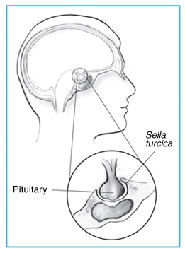
Sella turcica and pituitary gland.
Details
Identifiers
Latin sella turcica
MeSH D012658
TA98 A02.1.05.006
TA2 589
FMA 54709
Anatomical terms of bone

The sella turcica (Latin for 'Turkish saddle') is a saddle-shaped depression in the body of the sphenoid bone of the human skull and of the skulls of other hominids including chimpanzees, gorillas and orangutans. It serves as a cephalometric landmark. The pituitary gland or hypophysis is located within the most inferior aspect of the sella turcica, the hypophyseal fossa.

Contents

Structure

The sella turcica is located in the sphenoid bone behind the chiasmatic groove and the tuberculum sellae. It belongs to the middle cranial fossa. [1]

The sella turcica's most inferior portion is known as the hypophyseal fossa (the "seat of the saddle"), and contains the pituitary gland (hypophysis). In front of the hypophyseal fossa is the tuberculum sellae.

Completing the formation of the saddle posteriorly is the dorsum sellae, which is continuous with the clivus, inferoposteriorly. The dorsum sellae is terminated laterally by the posterior clinoid processes.

Development

It is widely believed that the development of the diaphragma sellae is a factor which determines the morphology of the sella turcica and its contents. [2]

Function

The sella turcica forms a bony seat for the pituitary gland.

Clinical significance

Empty sella syndrome is the condition of a shrunken or flattened pituitary gland.

Since the sella turcica forms a bony caudal border for the pituitary gland, a pituitary tumor usually extends upward in the rostral direction into the suprasellar region. This can result in compression of the optic chiasm, which lies on top of the pituitary, enveloping the pituitary stalk. Compression of the optic chiasm can lead to bitemporal hemianopsia, and, when there is no relevant trauma, this clinical finding is pathognomonic for a pituitary tumor.

Some pituitary adenomas can extend inferiorly, growing downward and invading the sphenoid bone and cavernous sinus. [3] Large adenomas can cause remodeling of the underlying sphenoid bone altering the shape of the sella turcica.[ citation needed ]

Sella turcica is also usually used as a reference point with nasion to establish the base of the skull in cephalometric analysis. This is commonly done prior to orthodontic treatment. [4]

Etymology

Sella turcica is from the Latin words sella, meaning seat, and turcica, meaning Turkish.

See also

Additional images

Related Research Articles

Articles related to anatomy include:

<span class="mw-page-title-main">Sphenoid bone</span> Bone at the front of the skull

The sphenoid bone is an unpaired bone of the neurocranium. It is situated in the middle of the skull towards the front, in front of the basilar part of the occipital bone. The sphenoid bone is one of the seven bones that articulate to form the orbit. Its shape somewhat resembles that of a butterfly or bat with its wings extended.

<span class="mw-page-title-main">Internal carotid artery</span> Artery of the human brain

The internal carotid artery is an artery in the neck which supplies the anterior circulation of the brain.

<span class="mw-page-title-main">Orbit (anatomy)</span> Cavity or socket of the skull in which the eye and its appendages are situated

In anatomy, the orbit is the cavity or socket/hole of the skull in which the eye and its appendages are situated. "Orbit" can refer to the bony socket, or it can also be used to imply the contents. In the adult human, the volume of the orbit is 30 millilitres, of which the eye occupies 6.5 ml. The orbital contents comprise the eye, the orbital and retrobulbar fascia, extraocular muscles, cranial nerves II, III, IV, V, and VI, blood vessels, fat, the lacrimal gland with its sac and duct, the eyelids, medial and lateral palpebral ligaments, cheek ligaments, the suspensory ligament, septum, ciliary ganglion and short ciliary nerves.

<span class="mw-page-title-main">Posterior cranial fossa</span> Area of the cranium containing the brainstem and cerebellum

The posterior cranial fossa is the part of the cranial cavity located between the foramen magnum, and tentorium cerebelli. It is formed by the sphenoid bones, temporal bones, and occipital bone. It lodges the cerebellum, and parts of the brainstem.

<span class="mw-page-title-main">Cavernous sinus</span> Sinus in the human head

The cavernous sinus within the human head is one of the dural venous sinuses creating a cavity called the lateral sellar compartment bordered by the temporal bone of the skull and the sphenoid bone, lateral to the sella turcica.

<span class="mw-page-title-main">Sphenoid sinus</span> One of the four paired paranasal sinuses

The sphenoid sinus is a paired paranasal sinus occurring within the body of the sphenoid bone. It represents one pair of the four paired paranasal sinuses. The pair of sphenoid sinuses are separated in the middle by a septum of sphenoid sinuses. Each sphenoid sinus communicates with the nasal cavity via the opening of sphenoidal sinus. The two sphenoid sinuses vary in size and shape, and are usually asymmetrical.

<span class="mw-page-title-main">Middle cranial fossa</span>

The middle cranial fossa is formed by the sphenoid bones, and the temporal bones. It lodges the temporal lobes, and the pituitary gland. It is deeper than the anterior cranial fossa, is narrow medially and widens laterally to the sides of the skull. It is separated from the posterior cranial fossa by the clivus and the petrous crest.

<span class="mw-page-title-main">Tuberculum sellae</span>

The tuberculum sellae is a slight median elevation upon the superior aspect of the body of sphenoid bone at the anterior boundary of the sella turcica and posterior boundary of the chiasmatic groove. A middle clinoid process flanks the tuberculum sellae on either side.

<span class="mw-page-title-main">Posterior clinoid processes</span>

The posterior clinoid processes are the tubercles of the sphenoid bone situated at the superior angles of the dorsum sellæ which represents the posterior boundary of the sella turcica. They vary considerably size and form. The posterior clinoid processes deepen the sella turcica, and give attachment to the tentorium cerebelli, and the dura forming the floor of the hypophyseal fossa.

<span class="mw-page-title-main">Anterior clinoid process</span>

The anterior clinoid process is a posterior projection of the sphenoid bone at the junction of the medial end of either lesser wing of sphenoid bone with the body of sphenoid bone. The bilateral processes flank the sella turcica anteriorly.

Pituitary apoplexy is bleeding into or impaired blood supply of the pituitary gland. This usually occurs in the presence of a tumor of the pituitary, although in 80% of cases this has not been diagnosed previously. The most common initial symptom is a sudden headache, often associated with a rapidly worsening visual field defect or double vision caused by compression of nerves surrounding the gland. This is often followed by acute symptoms caused by lack of secretion of essential hormones, predominantly adrenal insufficiency.

<span class="mw-page-title-main">Diaphragma sellae</span>

The diaphragma sellae or sellar diaphragm is a small, circular sheet of dura mater forming an (incomplete) roof over the sella turcica and covering the pituitary gland lodged therein. The diaphragma sellae forms a central opening to accommodate the passage of the pituitary stalk (infundibulum) which interconnects the pituitary gland and the hypothalamus.

<span class="mw-page-title-main">Body of sphenoid bone</span>

The body of the sphenoid bone, more or less cubical in shape, is hollowed out in its interior to form two large cavities, the sphenoidal sinuses, which are separated from each other by a septum.

<span class="mw-page-title-main">Chiasmatic groove</span> Groove whose anterior border is a ridge bounding the sphenoid bone

The chiasmatic groove is a transverse groove upon the superior aspect of the body of sphenoid bone within the middle cranial fossa. It is bounded anteriorly by the sphenoidal limbus, and posteriorly by the tuberculum sellae. The opening of each optic canal is placed at either lateral end of the chiasmatic sulcus. The optic chiasm is situated superior and quite posterior to the chiasmatic groove.

<span class="mw-page-title-main">Middle clinoid process</span>

The middle clinoid process is a small, bilaterally paired elevation on either side of the tuberculum sellae, at the anterior boundary of the sella turcica. A (larger) anterior clinoid process is situated lateral to each middle clinoid process. The diaphragma sellae and the dura of the floor of the hypophyseal fossa attach onto the middle clinoid processes.

<span class="mw-page-title-main">Chiasmal syndrome</span> Set of signs and symptoms that are associated with lesions of the optic chiasm

Chiasmal syndrome is the set of signs and symptoms that are associated with lesions of the optic chiasm, manifesting as various impairments of the affected's visual field according to the location of the lesion along the optic nerve. Pituitary adenomas are the most common cause; however, chiasmal syndrome may be caused by cancer, or associated with other medical conditions such as multiple sclerosis and neurofibromatosis.

<span class="mw-page-title-main">Base of skull</span> Inferior area of the skull, composed of the endocranium and lower parts of the skull roof

The base of skull, also known as the cranial base or the cranial floor, is the most inferior area of the skull. It is composed of the endocranium and the lower parts of the calvaria.

In anatomy, a fossa is a depression or hollow usually in a bone, such as the hypophyseal fossa. Some examples include:

Endoscopic endonasal surgery is a minimally invasive technique used mainly in neurosurgery and otolaryngology. A neurosurgeon or an otolaryngologist, using an endoscope that is entered through the nose, fixes or removes brain defects or tumors in the anterior skull base. Normally an otolaryngologist performs the initial stage of surgery through the nasal cavity and sphenoid bone; a neurosurgeon performs the rest of the surgery involving drilling into any cavities containing a neural organ such as the pituitary gland. The use of endoscope was first introduced in Transsphenoidal Pituitary Surgery by R Jankowsky, J Auque, C Simon et al. in 1992 G.

References

  1. Mancall, Elliott L.; Brock, David G., eds. (2011). "Cranial Fossae". Gray's Clinical Anatomy. Elsevier Health Sciences. p. 154. ISBN   9781437735802.
  2. Ferreri, A J M; Garrido, S A; Markarian, M G; Yañez, A (September 1992). "Relationship between the development of diaphragma sellae and the morphology of the sella turcica and its content". Surgical and Radiologic Anatomy. 14 (3): 233–239. doi:10.1007/BF01794946. PMID   1440188. S2CID   32329369.
  3. Knosp, Engelbert; Steiner, Erich; Kitz, Klaus; Matula, Christian (October 1993). "Pituitary Adenomas with Invasion of the Cavernous Sinus Space: A Magnetic Resonance Imaging Classification Compared with Surgical Findings". Neurosurgery. 33 (4): 610–618. doi:10.1227/00006123-199310000-00008. PMID   8232800.
  4. Proffit, William R. Contemporary Orthodontics, 4th Edition. C.V. Mosby, 122006. 6.5.2.1). vbk:978-0-323-04046-4#outline(6.5.2.1)