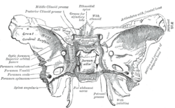
The sphenoid bone is an unpaired bone of the neurocranium. It is situated in the middle of the skull towards the front, in front of the basilar part of the occipital bone. The sphenoid bone is one of the seven bones that articulate to form the orbit. Its shape somewhat resembles that of a butterfly or bat with its wings extended.

The internal carotid artery is an artery in the neck which supplies the anterior circulation of the brain.

The sella turcica is a saddle-shaped depression in the body of the sphenoid bone of the human skull and of the skulls of other hominids including chimpanzees, gorillas and orangutans. It serves as a cephalometric landmark. The pituitary gland or hypophysis is located within the most inferior aspect of the sella turcica, the hypophyseal fossa.

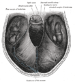
The posterior cranial fossa is the part of the cranial cavity located between the foramen magnum, and tentorium cerebelli. It is formed by the sphenoid bones, temporal bones, and occipital bone. It lodges the cerebellum, and parts of the brainstem.

In anatomy, a process is a projection or outgrowth of tissue from a larger body. For instance, in a vertebra, a process may serve for muscle attachment and leverage, or to fit, with another vertebra. The word is also used at the microanatomic level, where cells can have processes such as cilia or pedicels. Depending on the tissue, processes may also be called by other terms, such as apophysis, tubercle, or protuberance.

The pterygoid processes of the sphenoid, one on either side, descend perpendicularly from the regions where the body and the greater wings of the sphenoid bone unite.
The greater wing of the sphenoid bone, or alisphenoid, is a bony process of the sphenoid bone, positioned in the skull behind each eye. There is one on each side, extending from the side of the body of the sphenoid and curving upward, laterally, and backward.
The lesser wings of the sphenoid or orbito-sphenoids are two thin triangular plates, which arise from the upper and anterior parts of the body, and, projecting lateralward, end in sharp points [Fig. 1].

The squamous part of temporal bone, or temporal squama, forms the front and upper part of the temporal bone, and is scale-like, thin, and translucent.

The middle cranial fossa is formed by the sphenoid bones, and the temporal bones. It lodges the temporal lobes, and the pituitary gland. It is deeper than the anterior cranial fossa, is narrow medially and widens laterally to the sides of the skull. It is separated from the posterior cranial fossa by the clivus and the petrous crest.
The tuberculum sellae is a slight median elevation upon the superior aspect of the body of sphenoid bone at the anterior boundary of the sella turcica and posterior boundary of the chiasmatic groove. A middle clinoid process flanks the tuberculum sellae on either side.
The posterior clinoid processes are the tubercles of the sphenoid bone situated at the superior angles of the dorsum sellæ which represents the posterior boundary of the sella turcica. They vary considerably size and form. The posterior clinoid processes deepen the sella turcica, and give attachment to the tentorium cerebelli, and the dura forming the floor of the hypophyseal fossa.

The anterior clinoid process is a posterior projection of the sphenoid bone at the junction of the medial end of either lesser wing of sphenoid bone with the body of sphenoid bone. The bilateral processes flank the sella turcica anteriorly.
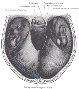
The diaphragma sellae or sellar diaphragm is a small, circular sheet of dura mater forming an (incomplete) roof over the sella turcica and covering the pituitary gland lodged therein. The diaphragma sellae forms a central opening to accommodate the passage of the pituitary stalk (infundibulum) which interconnects the pituitary gland and the hypothalamus.
The petrosal process is a sharp process below the notch for the passage of the abducent nerve on either side of the dorsum sellae of the sphenoid bone. It articulates with the apex of the petrous portion of the temporal bone, and forms the medial boundary of the foramen lacerum.

The body of the sphenoid bone, more or less cubical in shape, is hollowed out in its interior to form two large cavities, the sphenoidal sinuses, which are separated from each other by a septum.

The clivus or Blumenbach clivus is a bony part of the cranium at the base of the skull. It is a shallow depression behind the dorsum sellae of the sphenoid bone. It slopes gradually to the anterior part of the basilar occipital bone at its junction with the sphenoid bone. It extends to the foramen magnum. It is related to the pons and the abducens nerve.
The chiasmatic groove is a transverse groove upon the superior aspect of the body of sphenoid bone within the middle cranial fossa. It is bounded anteriorly by the sphenoidal limbus, and posteriorly by the tuberculum sellae. The opening of each optic canal is placed at either lateral end of the chiasmatic sulcus. The optic chiasm is situated superior and quite posterior to the chiasmatic groove.
The middle clinoid process is a small, bilaterally paired elevation on either side of the tuberculum sellae, at the anterior boundary of the sella turcica. A (larger) anterior clinoid process is situated lateral to each middle clinoid process. The diaphragma sellae and the dura of the floor of the hypophyseal fossa attach onto the middle clinoid processes.

The base of skull, also known as the cranial base or the cranial floor, is the most inferior area of the skull. It is composed of the endocranium and the lower parts of the calvaria.